Digital Facial Approximation and Digital Reconstruction of the Projectile Trajectory in Charles XII of Sweden: A 3D Biomechanical and Craniometric Analysis
Highlights
● First quantitative cephalometric analysis of Charles XII reveals Class III maxillary retrognathism and mandibular prognathism, providing craniometric validation for features observed in historical portraits and the death mask.
● First complete 3D anatomical reconstruction integrating skull, brain voxel data, and intracranial venous system of Charles XII, enabling precise identification of structures damaged by the projectile.
● Spatial verification of the tricorn hat against the documented intracranial trajectory — a geometrical check absent from prior literature — confirms geometric compatibility with the left temporal entry point.
● Cranial reconstruction achieved exclusively from 1917 autopsy photographs and radiographs via anatomical deformation, demonstrating OrtogOnBlender’s applicability to cases with no direct 3D scan available.
● Forensic facial approximation of Charles XII produced using open-source tools, converging with portrait evidence and death mask morphology through independent anatomical projections.
Abstract
This study presents an unprecedented digital bioarchaeological and forensic analysis of Charles XII of Sweden, whose death in 1718 remains one of Europe’s greatest historical mysteries. Utilizing open-source technologies, specifically the OrtogOnBlender ecosystem, the research performed a forensic facial approximation of the monarch and a 3D reconstruction of the fatal projectile’s trajectory. In the absence of a direct 3D scan of the skull, the skeletal structure was reconstructed through anatomical deformation by cross-referencing metric data from photographs and radiographs taken during the 1917 exhumation. The results precisely corroborated the historical data from the last autopsy, confirming a nearly rectilinear and horizontal transfixing trajectory, with entry through the left temporal region and exit through the right. Innovatively, the study applied a USP standard cephalometric analysis, revealing a clinical condition of maxillary retrognathism associated with mandibular prognathism, which explains the projected lower lip physiognomy observed in portraits and the death mask. In addition to the face, the brain and vascular structures were modeled, allowing for the identification of the injured encephalic areas. Finally, the integration of the tricorne hat into the digital model demonstrated exact geometric compatibility with the ballistic path, filling gaps in previous studies and consolidating an efficient methodology for the analysis of complex historical cases.
Keywords: Charles XII of Sweden, Forensic Facial Approximation, 3D Ballistic Trajectory, Cephalometry, Anatomical Reconstruction, OrtogOnBlender
Introduction
Brief Biography of Charles XII
Charles XII was born in Stockholm on June 17, 1682, the son of Charles XI and Ulrika Eleonora of Denmark. After his mother’s death in 1693, he developed a very close relationship with his father, accompanying him on official inspections and travels. Following Charles XI’s death in 1697, he was declared of age and crowned at just 15 years old, becoming the last absolute monarch of Sweden. Described as a ruler of the early Enlightenment era, Charles XII was known for his asceticism and radical values. He was deeply committed to duty, possessed a solid education in theology and languages, and demonstrated remarkable shrewdness both as a statesman and as a general. His 21-year reign was defined by the defense of Sweden during the Great Northern War. Initially, he achieved brilliant victories (such as at Narva in 1700) and was regarded as a skilled tactician. However, the invasion of Russia (1707–1709) proved disastrous. He faced a scorched-earth strategy — the same one that would later weaken Napoleon and Hitler — along with the harsh Russian winter, culminating in the defeat at Poltava (1709), which destroyed the Swedish army and ended Sweden’s status as a great power. After the defeat, he lived in forced exile for five years in the Ottoman Empire (present-day Moldova/Turkey), from where he continued attempting to influence European politics. He returned to Sweden in 1714, finding the empire in decline, yet still promoted significant domestic reforms and administrative measures that were ahead of their time. His life ended abruptly at the age of 36 on November 30, 1718, during the siege of the Fredrikshald fortress (in present-day Norway). He was struck in the temple by a projectile while inspecting the trenches — an event that marked the end of Swedish hegemony in the Baltic and remains one of history’s greatest mysteries, as it is still debated whether the fatal shot was fired by an enemy or came from his own ranks (friendly fire) [Hatton_and_Nordlund_2026_e].
The Autopsies
Three autopsies were performed on the body of Charles XII: one in 1746, another in 1859, and the final one in 1917 [Junno_et_al_2022_e] [Key-Aberg_and_Stille_1918_e].
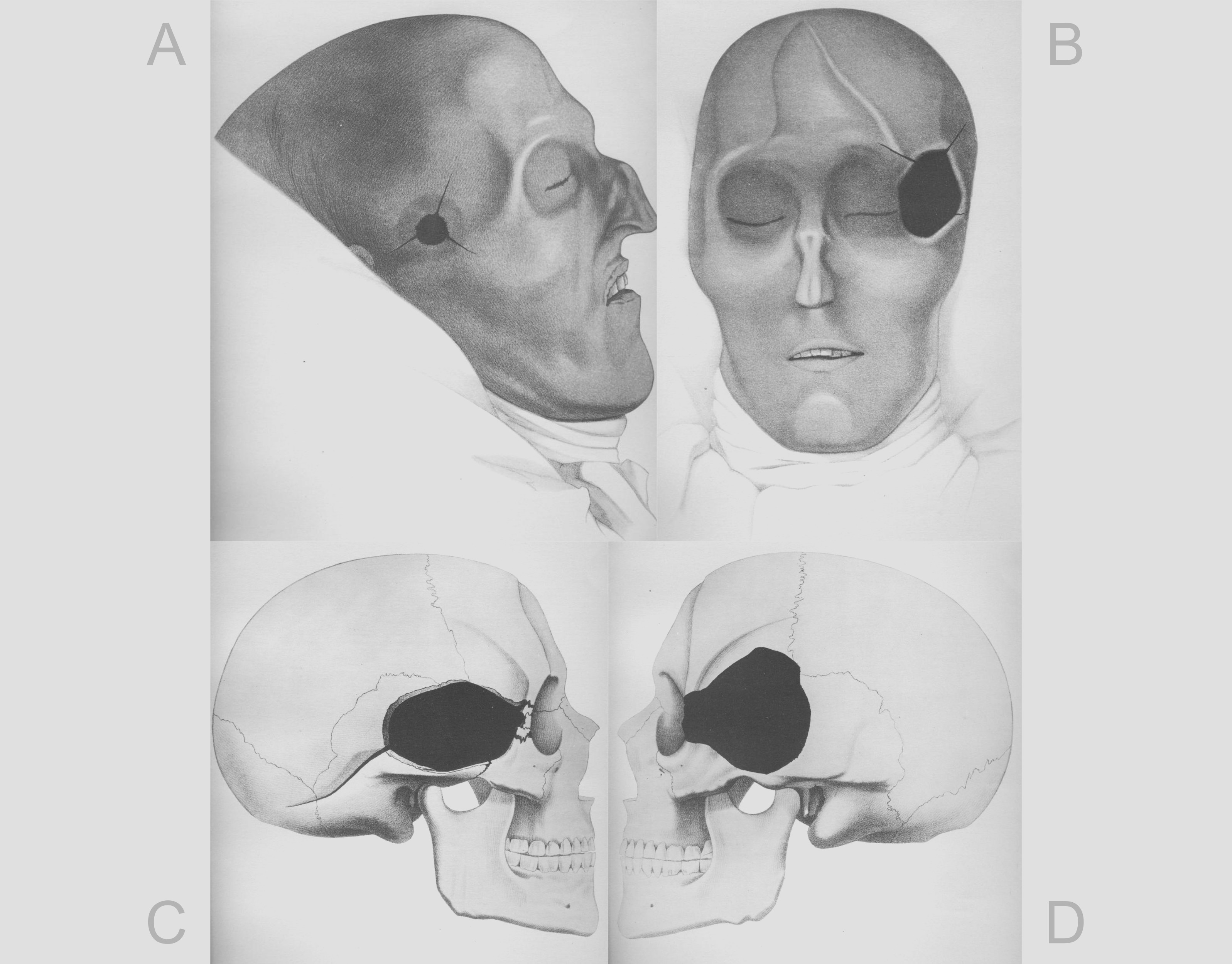
Fig. 39 Engravings from the 1859 autopsy. A) Soft tissue wound on the King’s right temple. B) Soft tissue wound on the King’s left temple. C) Representation on a surrogate skull (not the King’s) of the cranial damage on the right side. D) Representation on a surrogate skull of the cranial damage on the left side. Public domain images. Digitization available at: https://brf-portalen.se/wp-dragoner/album/k12-banesar/index.html
The 1859 results indicated that the head wound was fatal, resulting from a firearm discharge that would have caused instantaneous death. The projectile entered through the left side, at the outer edge of the bony ring of the eye socket, and emerged slightly in front of the right ear in a nearly horizontal trajectory. Although uncertainties remained regarding the projectile type, it was considered more likely to be a musket ball or a canister shot, and less likely to be a piece of scrap metal or a bomb fragment. Based on the distance it traveled after transfixing the King’s head, it was estimated that the projectile was already decelerating at the moment of impact. The investigation evidenced that it was a lesion caused by a single projectile, with no residues found inside the skull [Key-Aberg_and_Stille_1918_e]. The examination relied on manual illustrations of the King’s head and utilized a surrogate skull as a didactic element to project the damage onto the bone tissue (Fig. 39).
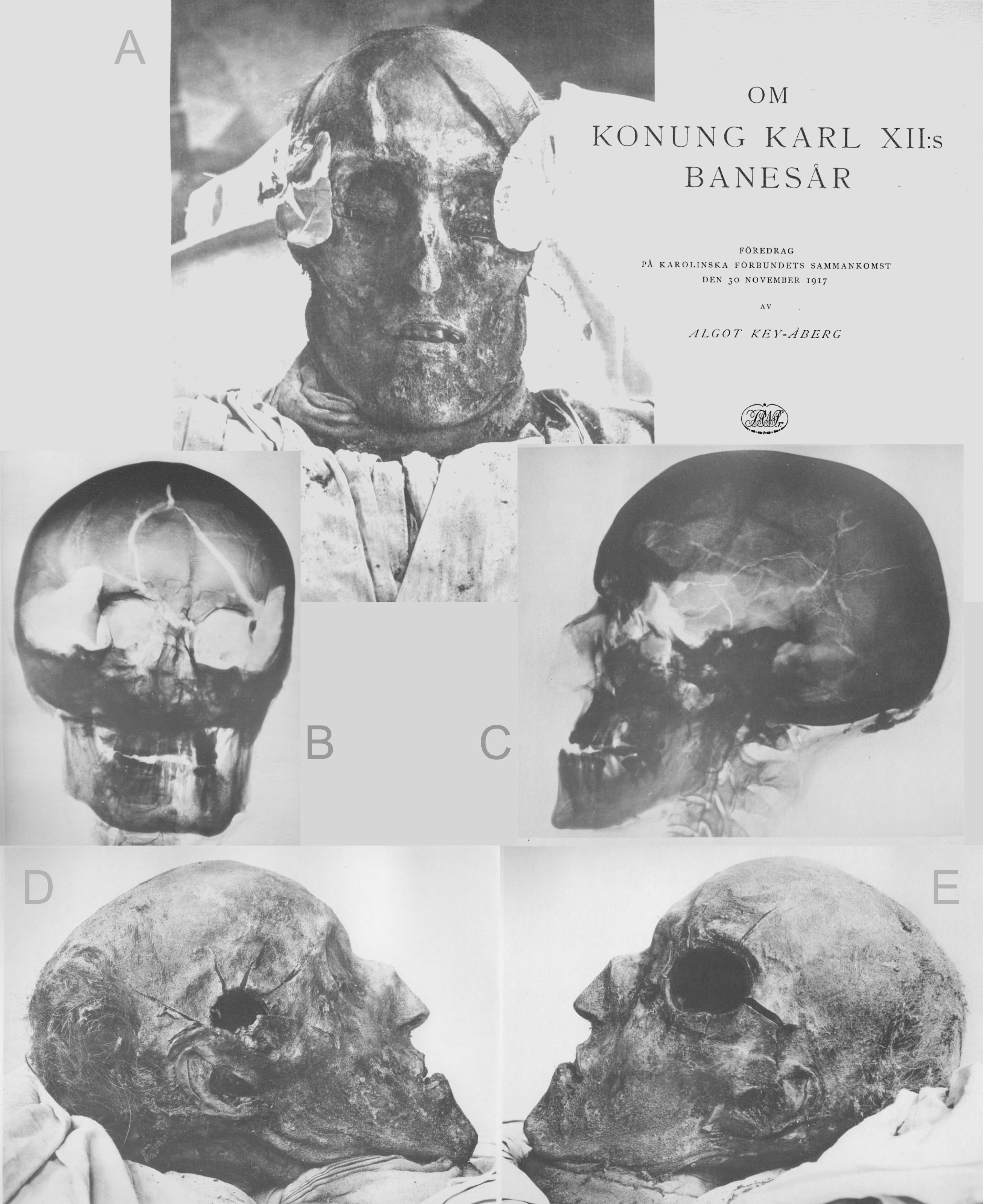
Fig. 40 Photographs and radiographs from the 1917 autopsy. A) Frontal view of the King’s head with dressings still applied and the cover of the 1917 autopsy book, published in 1918. B) Radiograph of the King’s head (frontal view). C) Radiograph of the left lateral portion of the partially reconstructed skull. D) Photographic image of the soft tissue wound on the right temple. E) Photographic image of the soft tissue wound on the left temple. Public domain images. Digitization available at: https://brf-portalen.se/wp-dragoner/album/k12-banesar/index.html
The 1917 autopsy, unlike the previous one which relied solely on manual illustrations, utilized technologies such as photographs and radiographs. This examination confirmed prior findings and expanded the analysis with additional data. Specialists observed that the King’s death was instantaneous, caused by a single projectile. The entry point was located in the anterior part of the left temporal region, with the exit on the right, in the anterior half of the squamous part of the temporal bone. The projectile did not deviate, maintaining a straight trajectory slightly oriented toward the back, with no significant inclination relative to the horizontal plane. The projectile was proven to have a circular cross-section and a spherical shape, with an estimated diameter between 18 and 20 mm, allowing for a variation of one or two millimeters (16 to 22 mm). The examination provided no evidence that the shot was fired at close range. It was emphasized that the current shape of the lesion could not serve as a parameter for the original appearance of the opening, as it had been influenced by subsequent interventions, primarily the King’s embalming process. It was concluded that the projectile transfixed the head with high kinetic energy [Key-Aberg_and_Stille_1918_e].
Subsequent Publications
Several books have been written about the case and, currently, two peer-reviewed articles stand out in their approach. One of them, published in Forensic Science International [Nordling_1998_e], addresses the general context, presenting a mosaic of research over time. The author, while indicating that the event could be a regicide, also provides data that limits this claim, such as the fact that Charles XII constantly exposed himself to enemy fire and, on the night of his passing, 22 other soldiers were also struck.
Another relevant study was published in 2022 in the journal PNAS Nexus [Junno_et_al_2022_e]. In this work, the authors concluded that the projectile could not have been made of lead nor possessed a diameter of 19.5 mm—corresponding to the mark on the felt hat worn by the monarch—but was instead a caliber exceeding 20 mm. The researchers mention that, at the time, iron canister shot measured 21.9 mm, indicating material and structural compatibility. To support these conclusions, ballistic tests were conducted on skulls filled with gelatin and on a folded piece of felt.
Curiously, the 1917 study also performed ballistic tests with a dry skull and another filled with gelatin and, as previously mentioned, already considered a potential diameter of 22 mm [Key-Aberg_and_Stille_1918_e], which accommodates the 21.9 mm cited in the modern study. Complementary information will be explored throughout the text in the Results and Discussion section.
Scope of the Present Study
The objective of the present study is to perform a forensic facial approximation that will provide a cranial structure closest to that of King Charles XII. Based on this, it aims to reconstruct/approximate the brain structure and local veins, thereby defining the trajectory of the projectile and the potentially injured regions.
Note
The analysis presented here adds to a series of other historical approaches related to facial approximation and anatomical analysis, all with a didactic purpose, aiming at the use of the OrtogOnBlender add-on and open-source software for technical collaborations that can reinforce previous studies or provide novel assessments.This collection includes the following studies: The Facial Approximation of a Battle of Gotland Victim (1361) [Moraes_et_al_2022_e], The Facial Approximation and Accident Dynamics of Phineas Gage (1848) [Moraes_2023_e], The Facial Approximation and Structural Analysis of the Piltdown Man Skull - Fraud (1912) [Moraes_Bezzi_and_Bezzi_2024_e], The Facial Approximation of the “Vampire” of Venice (15th-17th Century) [Moraes_2024_e], The Forensic Facial Approximation of Seqenenre Tao II (c. 1558-1553 BC) [Moraes_and_Habicht_2024_e], The Facial Approximation of the Porsmose Man (c. 5500 BP) [Moraes_et_al_2025_e], and 3D Digital Analysis of the Anatoli Bugorski Case (1978) [Moraes_and_Moura_2025].
Materials and Methods
Forensic facial approximation is a technique that consists of generating a face from the morphological elements present in the skull. The present work followed the protocol described in detail in [Moraes_et_al_2024_e] and, complementarily, in [Moraes_et_al_2023_e]. For the data preparation, free and open-source software was used, such as the OrtogOnBlender add-on (https://www.ciceromoraes.com.br/doc/pt_br/OrtogOnBlender/index.html), which enables the Blender software (https://www.blender.org/) as a workstation suite for projects applied to health sciences, as well as Inkscape (https://inkscape.org/) and Gimp (https://www.gimp.org/) for generating graphics, refining, and editing images. All tools were executed on the Linux Ubuntu 24.04 operating system (https://ubuntu.com/). Anatomical terms were consulted in [Tank_and_Gest_2009_e].
Fig. 41 A) Lateral and frontal projections of the skull. B) Anatomical deformation over the lateral projection. C) Resulting skull. D) Projection of anatomical structures. E) Soft tissue thickness markers and facial profile projection. F) Anatomical deformation and profile projection. G) Final bust mesh interpolating the generated data. H) Confluence of all data into a resulting face. I) Configured facial hair.
As a 3D digitized skull of King Charles XII is unavailable, the solution found to obtain it consisted of cross-referencing data from the photographs and radiographs present in [Key-Aberg_and_Stille_1918_e], grouping all the necessary information—from the general limits of the skull and key measurements to the position of the lesions caused by the projectile (Fig. 41, A). This cranial reconstruction technique followed the approach presented in [Moraes_et_al_2024_e]. With the frontal and lateral images, it became possible to model a skull or adjust the anatomy of a virtual donor using the anatomical deformation technique. It was decided to import a donor head composed not only of the skull but also of the soft tissue (face), adjusting it so that the donor’s bones were made compatible with the King’s bone structure (Fig. 41, B). The resulting skull (Fig. 41, C) received soft tissue thickness markers based on a sample of modern European males, with an average BMI and an age range of 30–39 years [De_Greef_2006_e]. Complementarily, anatomical points were positioned to generate structural projections based on tomographic measurements of living individuals [Moraes_and_Suharschi_2022_e], which provide anatomical boundaries for lips, eyes, nasal wings, ears, and other structures (Fig. 41, D). A lateral projection of the nose was carried out, also based on tomographic data from living people [Moraes_and_Suharschi_2022_e], which allowed tracing the facial profile in conjunction with the tissue thickness markers (Fig. 41, E). A minor adjustment was required on the mandible, which was slightly open; when overlaying the result of the anatomical deformation and the profile projection, the data proved to be confluent (Fig. 41, F). A mesh corresponding to the final bust was generated through the interpolation of all facial projections (Fig. 41, G). In frontal view, the anatomical limitations of the face proved to be compatible with the statistical projections previously performed (Fig. 41, H). The bust was refined via digital sculpting, in addition to the configuration of facial hair, whose references were extracted from portraits of Charles XII (Fig. 41, I). The hair was adjusted based on a portrait of the King dating from 1717, in which hair loss is significant compared to portraits from his youth (see Fig. 45, E).
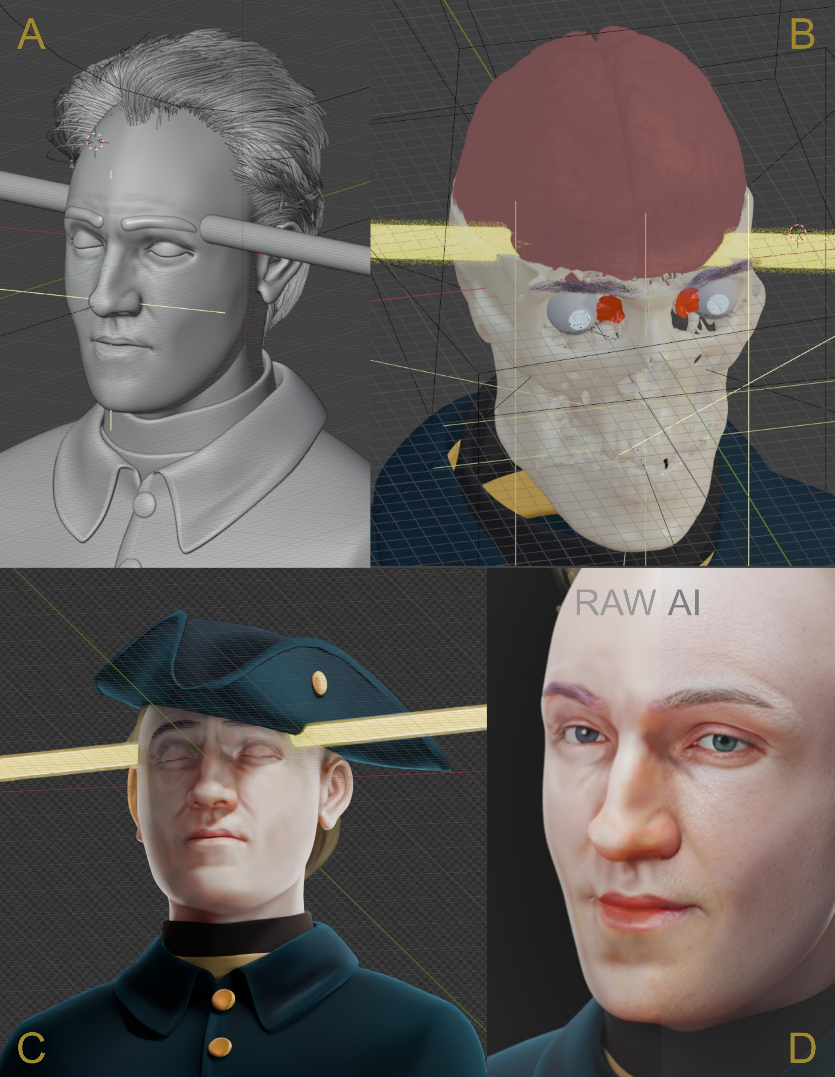
Fig. 42 A) Projectile tunnel crossing the final bust already with clothing. B) Adjusted brain voxel data. C) Rendering preview with the tricorne hat adjusted to the projectile entry. Raw rendering on the left and AI-enhanced adjustment on the right.
The bust received manual modeling of the clothing based on portraits of Charles XII and historical photography (https://commons.wikimedia.org/wiki/File:St%C3%B6vel_-_Livrustkammaren_-_8783.tif). Additionally, a cylinder of variable diameter, representing the potential projectile thicknesses, was modeled and adjusted to the lesions (Fig. 42, A). The skull received an overlay of anatomical information, such as the cerebral venous system (https://data.kitware.com/#collection/591086ee8d777f16d01e0724/folder/58a372e38d777f0721a64dc6) and the voxel data of a brain [Edlow_2019_e], properly adjusted to the endocranium (Fig. 42, B). A tricorne hat was imported from a model available for download on the Sketchfab portal (https://sketchfab.com/3d-models/tricorn-hat-lowpoly-ac0ecde570ba49c9adecd0b6820a84c0), provided under a Creative Commons license by user Aarte. The structure was adjusted to conform to the King’s head, strictly respecting the entry point of the projectile (Fig. 42, C). Only one image received localized treatment on the frontal portion of the face with detail enhancement via artificial intelligence, using a weight of 0.99 (on a scale of 0 to 1), ensuring high fidelity to the original and enriching the facial surface with fine details (Fig. 42, D). All other images did not use AI technology, and even the one that received such treatment underwent additional adjustments via image editing in Gimp.
Important
In the Data Statement section, video lessons detailing the aforementioned techniques—such as the projection of anatomical boundaries, nasal tracing, and anatomical deformation—will be made available. This content corroborates the references already available for download, in strict accordance with the open science approach adopted by the studies shared in OrtogOnLineMag. Such choices extend beyond mere rhetoric, as there are peer-reviewed publications that have employed the methodologies presented in the referred research, notably an article in Forensic Science International [Nasca_et_al_2026_e].
Results and Discussion
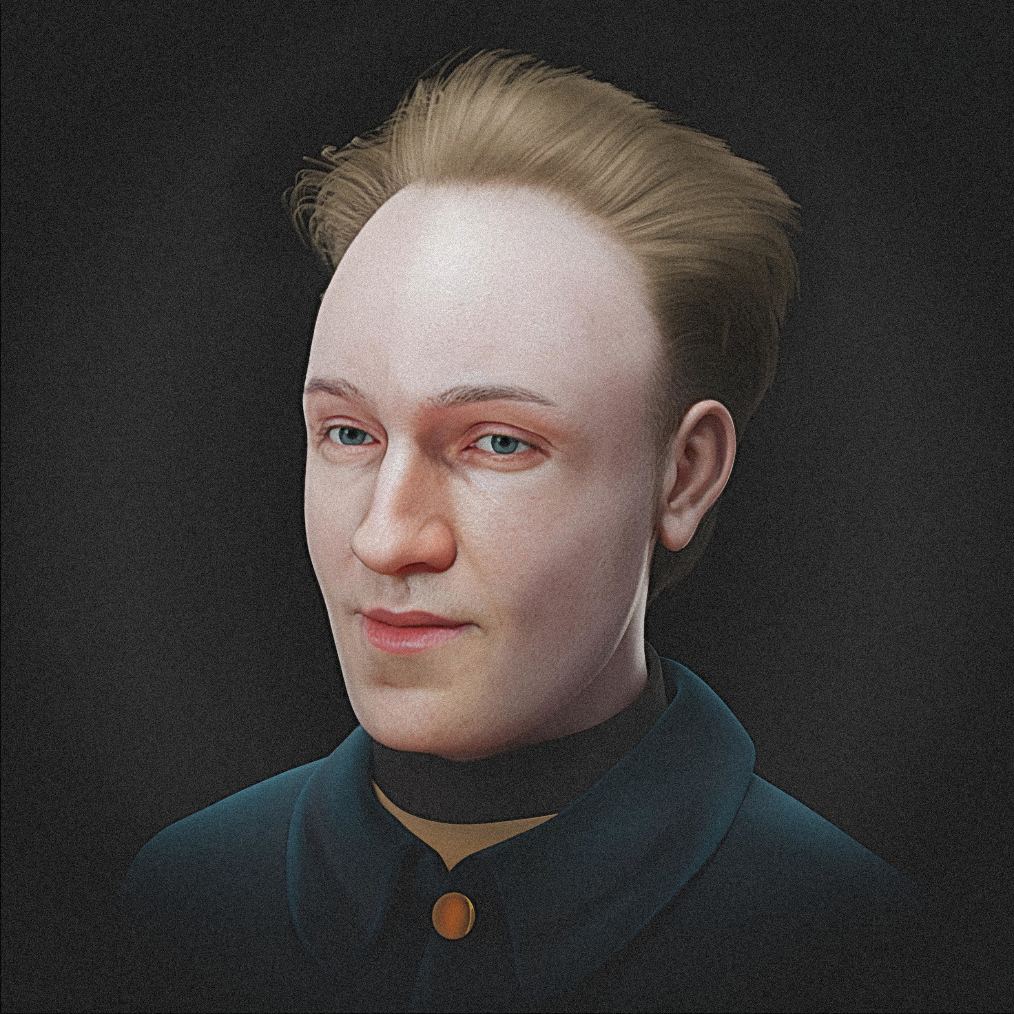
Fig. 43 Forensic facial approximation - final image.
The final image resulting from the facial approximation process contains objective elements derived from the skull and historical elements, along with subjective components from portraits, making it the only one to receive minimal retouching via AI (Fig. 43). The focus of the present work lies primarily on utilizing the final bust for the analysis of the projectile’s trajectory; however, the methodological standard followed the same technical rigor applied to historical faces in artistic contexts and to the faces of crime victims in a forensic scope [Baldasso_et_al_2020_e].
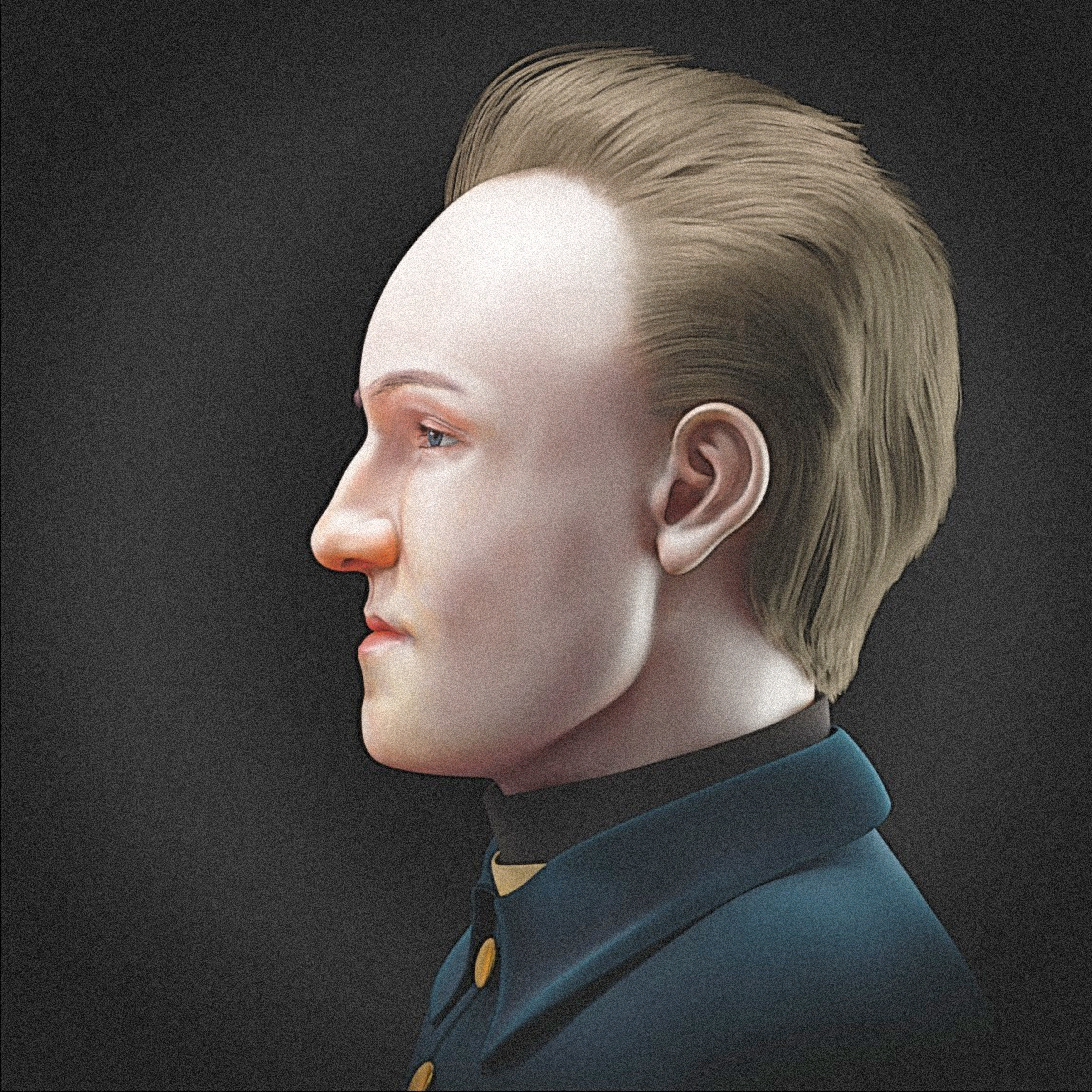
Fig. 44 Forensic facial approximation - profile view (with evident maxillary retrognathism and mandibular prognathism).
One aspect that immediately drew attention upon viewing the King’s lateral radiograph was the characteristic of potential maxillary retrognathism associated with mandibular prognathism, which became evident in the profile view (Fig. 44).
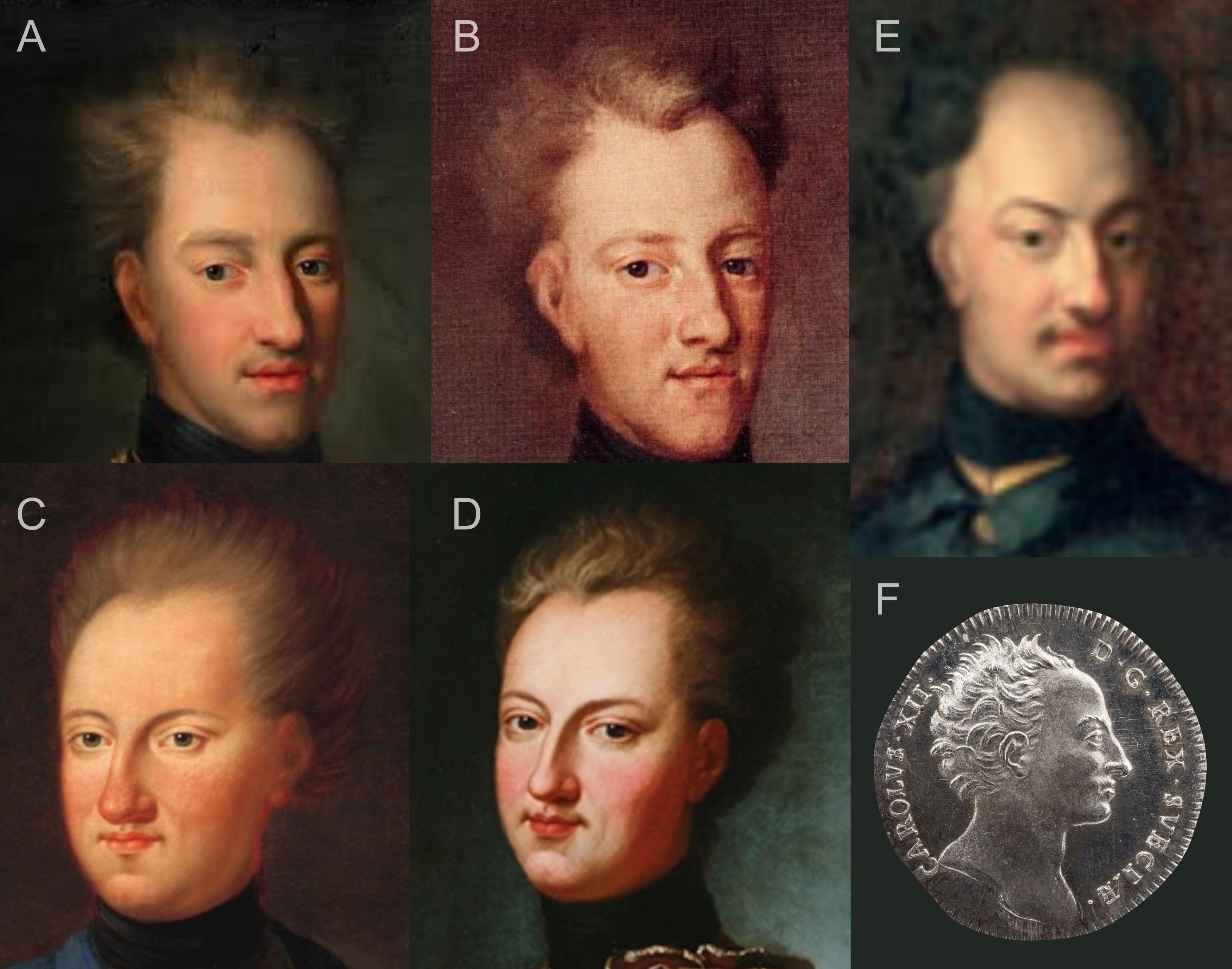
Fig. 45 A) Karl XII of Sweden. C.1700, Bema Knowledge (https://pt.wikipedia.org/wiki/Ficheiro:Karl_XII_of_Sweden._C.1700.jpg). B) Karl XII 1706 by David von Krafft (https://commons.wikimedia.org/wiki/File:Karl_XII_1706.jpg). C) Charles VII of Swedenby Axel Sparre (https://commons.wikimedia.org/wiki/File:Charles_VII_of_Sweden.jpg). D) Copy Charles XII - Nationalmuseum (https://en.wikipedia.org/wiki/File:Copy_Charles_XII_-_Nationalmuseum_-_17886.png). E) Karl XII i Lund by David von Krafft (https://commons.wikimedia.org/wiki/File:Karl_XII_i_Lund.jpg). F) Kastpenning med Karl XIIs profil, 1719, Miguel Herranz (https://commons.wikimedia.org/wiki/File:Kastpenning_med_Karl_XIIs_profil,_1719_-_Livrustkammaren_-_100562.tif).
These characteristics are also present in the portraits attributed to the King, whether those painted from life, copies, or even posthumous works. In addition to the characteristic hair and hairstyle, as well as the clothing replicated in this work, one can perceive a straight or slightly downward-slanting nose, a retracted supralabial region, and a projected lower lip, along with, occasionally, a prominent mentum (chin) (Fig. 45).
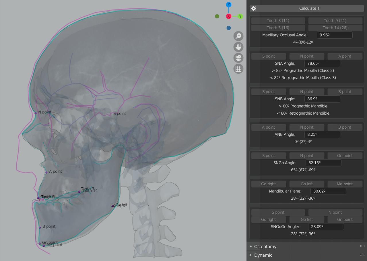
Fig. 46 USP Cephalometry in OrtogOnBlender.
To quantitatively evaluate the facial structure, a USP standard cephalometry [USP_2021_e] [Moraes_et_al_2023_e] was performed by positioning the relevant anatomical points (Fig. 46). The result was consistent with visual observations, as the SNA angle resulted in 78°40’—a value lower than 82°, which configures maxillary retrognathism (Class III). Furthermore, the SNB angle resulted in 86°54’ with the mandible in the resting position (according to the radiograph) and 88°29’ in occlusion; these values, being above 80°, configure mandibular prognathism.
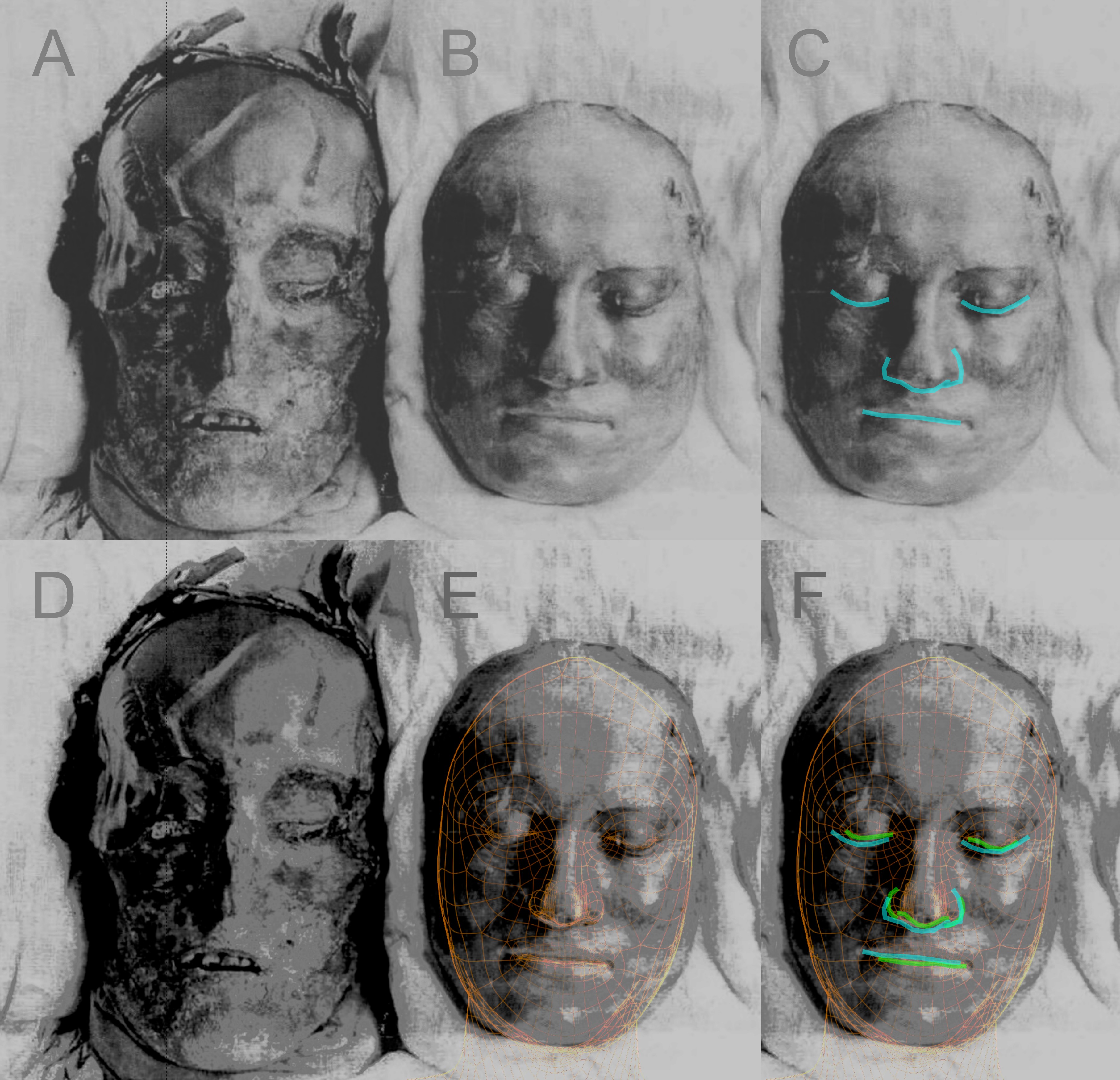
Fig. 47 Composition made from a photograph of King Charles XII’s body alongside his death mask (1917). Otto Mattsson (https://commons.wikimedia.org/wiki/File:CharlesXII_mummy_with_Death_mask.jpg).
Although portraits maintain the general characteristics attributed to Charles XII, the heterogeneity of these representations raises questions regarding their fidelity to the actual face. One way to circumvent this limitation is through the analysis of life or death masks. In the present study, a death mask was analyzed, starting with a comparison to a frontal photograph capturing the monarch’s body—already dehydrated after nearly 200 years—and his death mask laterally (Fig. 47, A and B). It is notable that the death mask presents a considerably robust face, not only in relation to the deceased body, which would be expected, but also when compared to the portraits. The method of the mask’s fabrication was not evaluated; however, the process, due to the pressure exerted and the characteristics of the body’s position and mandibular rotation, may have contributed to a slight deformation relative to the original, added to the trauma of death and the beginning of the natural decomposition process. Despite these variables, lines were traced at the corners of the eyes, nose, and mouth (Fig. 47, B) to compare them with the approximated face, which was overlaid in wireframe onto the photograph (Fig. 47, C). The subsequent comparison of the edges evidenced that the mask’s nose was slightly more downward-inclined and the labial commissure line was somewhat higher (Fig. 47, D), alterations compatible with the aforementioned deformations.
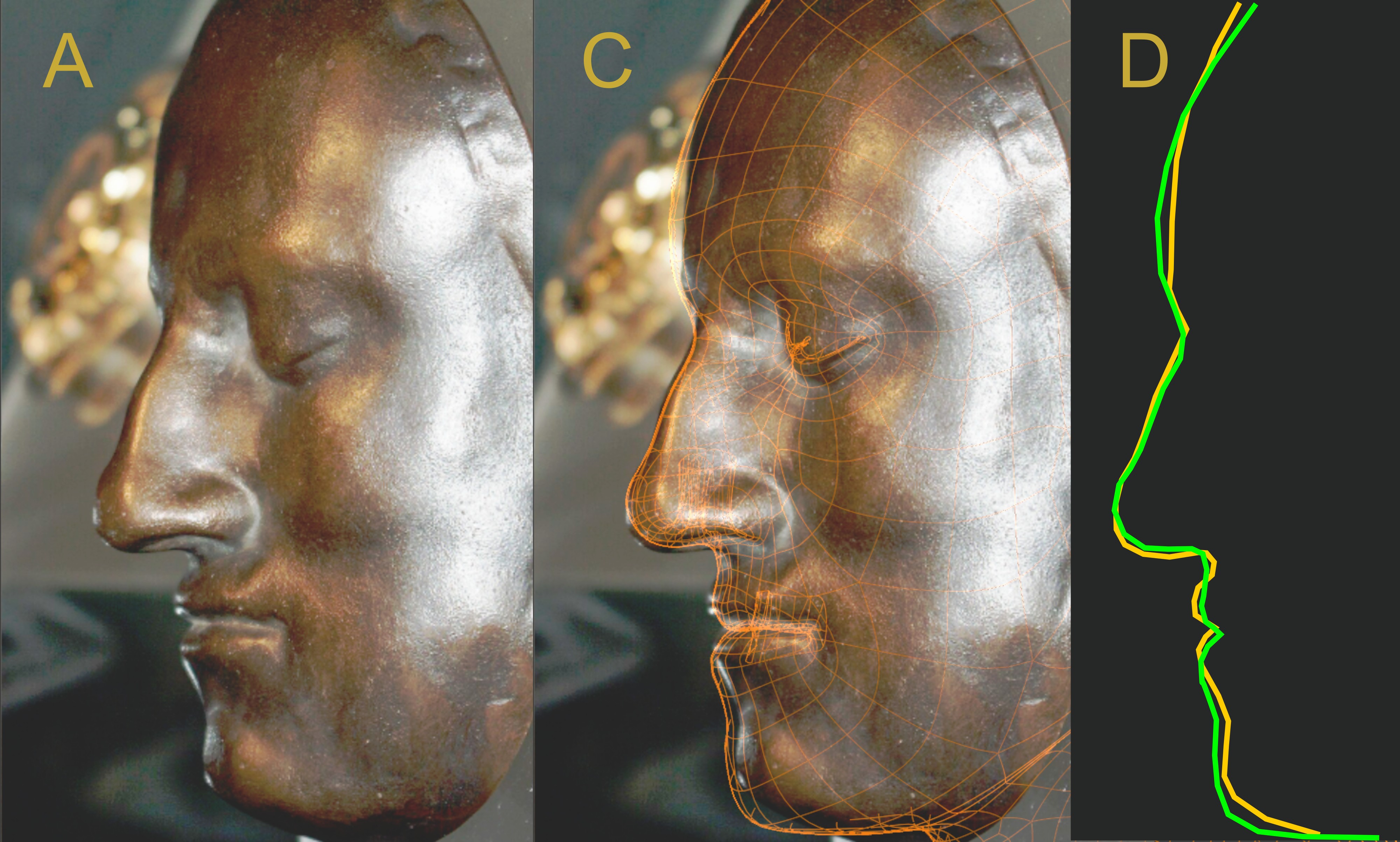
Fig. 48 A) Death mask in profile view. B) Overlay of the facial approximation. C) Comparison of the death mask’s profile (in yellow) relative to the facial approximation (in cyan). Sven Rosborn (https://commons.wikimedia.org/wiki/File:Karl_XIIs_d%C3%B6dsmask.jpg).
A closer, higher-quality photograph of the death mask captured in profile (Fig. 48, A) allowed for a more precise overlay with the facial approximation (Fig. 48, B). The procedure reinforced the findings from the frontal view: although the eyes were compatible, as was the nose in general and the height of the lips, the mask’s nose remains slightly more downward-inclined, while the lips are somewhat higher (Fig. 48, C). However, an evident feature is the projection effect of the upper lip—a common phenomenon in individuals with maxillary retrognathism during dental occlusion, where the labial mass is compressed, creating a projected appearance. This volume may indicate that the occlusion in the mask is more severe than that established in the cranial correction from the radiograph, a hypothesis supported by the difference in chin height, given that the chin of the facial approximation is positioned slightly below what is observed in the death mask. Nevertheless, due to the minor variation of the structures, the result is considered structurally satisfactory. The discrepancy in the forehead region could be indicative of various situations, ranging from local compression during the mask’s molding to the projection of soft tissue on a skull structurally compromised by the multiple lesions caused by the projectile.
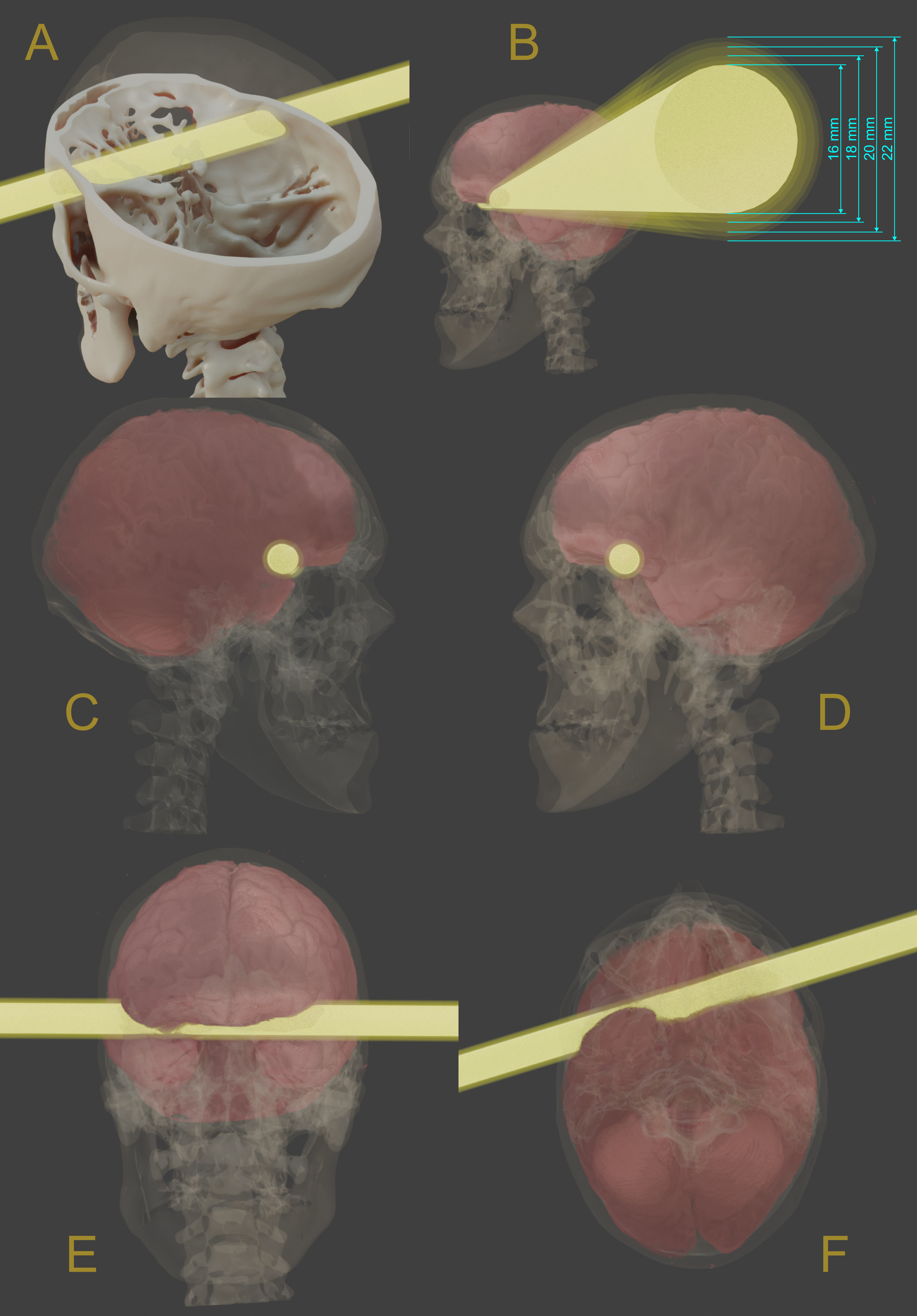
Fig. 49 A) Projectile path through the skull. B) Diameters encompassing the 1917 analysis. C) Visualization of the right side based on the projectile’s rotation. D) Visualization of the left side based on the projectile’s rotation. E) Frontal view of the projectile’s passage. F) Inferior view of the projectile’s passage.
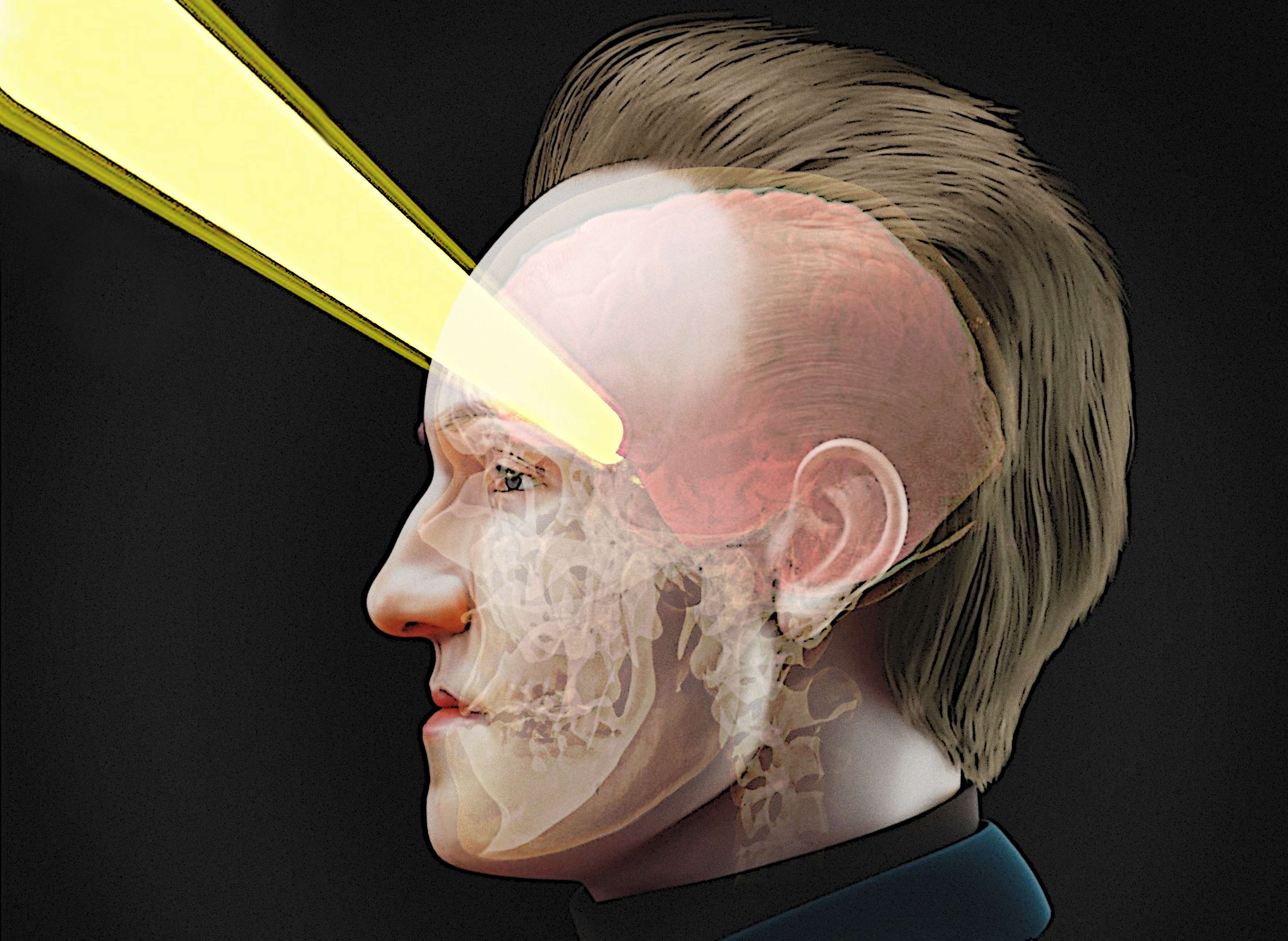
To trace the entry and exit of the projectile, the profile photographs from the 1917 exhumation with intact soft tissue were used as a reference (Fig. 40, D and E). As previously discussed, the specialists who performed the exhumation discarded the use of the skeletal lesion as a solid parameter for assessing the projectile’s thickness [Key-Aberg_and_Stille_1918_e]; therefore, the tracing was adjusted based on the center of the two soft tissue wounds. This procedure proved consistent with previous approaches, although minor alignment discrepancies may occur due to the margin of error and the use of donor skulls as references, as observed in [Nordling_1998_e] (Figures 5 and 6) and in KXII skalle rekonstrucktion av skottet (https://commons.wikimedia.org/wiki/File:Avgjutning_av_KXIIs_skalle_m_rekonstruktion_av_skottet_-_Livrustkammaren_-_27057.tif); in both cases involving a surrogate skull. The entry was observed between the greater wing of the sphenoid and the frontal process on the left side, with the exit through the right squamous part (Fig. 49, A). Using the parameters from the [Key-Aberg_and_Stille_1918_e] study, which stipulated a projectile with a diameter between 18 and 20 mm, with a margin of error of ±1 mm and ±2 mm, a tunnel was created with thicknesses of 16, 17, 18-20, 21, and 22 mm to encompass this data (Fig. 49, B). A virtual donor brain was adjusted to fit Charles XII’s endocranium, allowing for the observation of the affected regions (Fig. 49, C and D). From the left side, the tunnel hits the orbital part of the frontal lobe and involves the anterior portion of the right frontal lobe (Fig. 49, E and F).
One of the most intriguing features of Charles XII’s cranial lesions, widely documented in the 1917 exhumation report, is the presence of a large entry defect on the left side of the head, contrasting with a significantly smaller exit hole on the right. This pattern, the inverse of what is commonly observed in modern high-velocity projectile wounds, can cause confusion for readers accustomed to media representations of ballistic injuries. In contemporary projectiles—generally faster, smaller, and with greater gyroscopic stability—the exit wound is usually larger due to tumbling and higher energy transfer at the end of the trajectory [Kimmerle_and_Baraybar_e]. However, in the case of early 18th-century musket balls—bulky projectiles (approximately 18–22 mm) made of almost pure lead (or other metal) and fired at low velocities (typically between 200–400 m/s)—the behavior is different: the projectile tends to deform and flatten upon impacting the cortical bone at entry, creating a larger hole, while at the exit, already decelerated and deformed, it produces a smaller and more irregular defect. Recent ballistic experiments [Junno_et_al_2022_e] successfully reproduced this pattern by using projectiles similar to those available in 1718, reinforcing the forensic coherence of the 1917 observations, whose specialists also conducted such tests [Key-Aberg_and_Stille_1918_e].
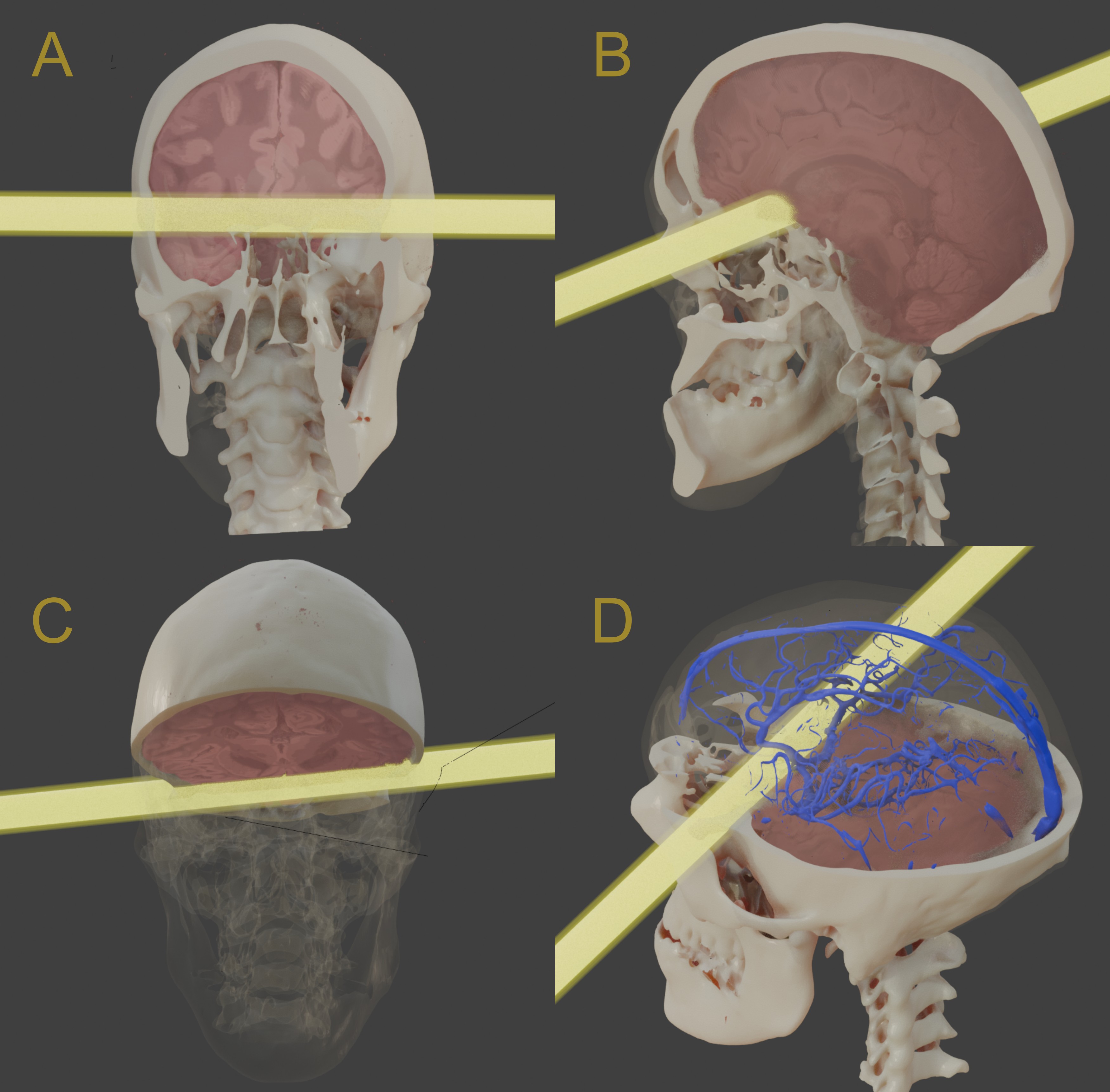
Fig. 50 Brain voxel data slices: A) Frontal view. B) Lateral view. C) Slightly inferior view. D) Superior view with the addition of cerebral veins.
When observed internally, it is evident that the trajectory transfixed the frontal and temporal lobes, potentially striking the olfactory tract and bulb (Fig. 50, A), the corpus callosum, and the septum pellucidum (Fig. 50, B and C). By reconstructing the vascular network of the region, it is observed that the projectile could have hit, among other structures, the middle cerebral artery (Fig. 50, D).
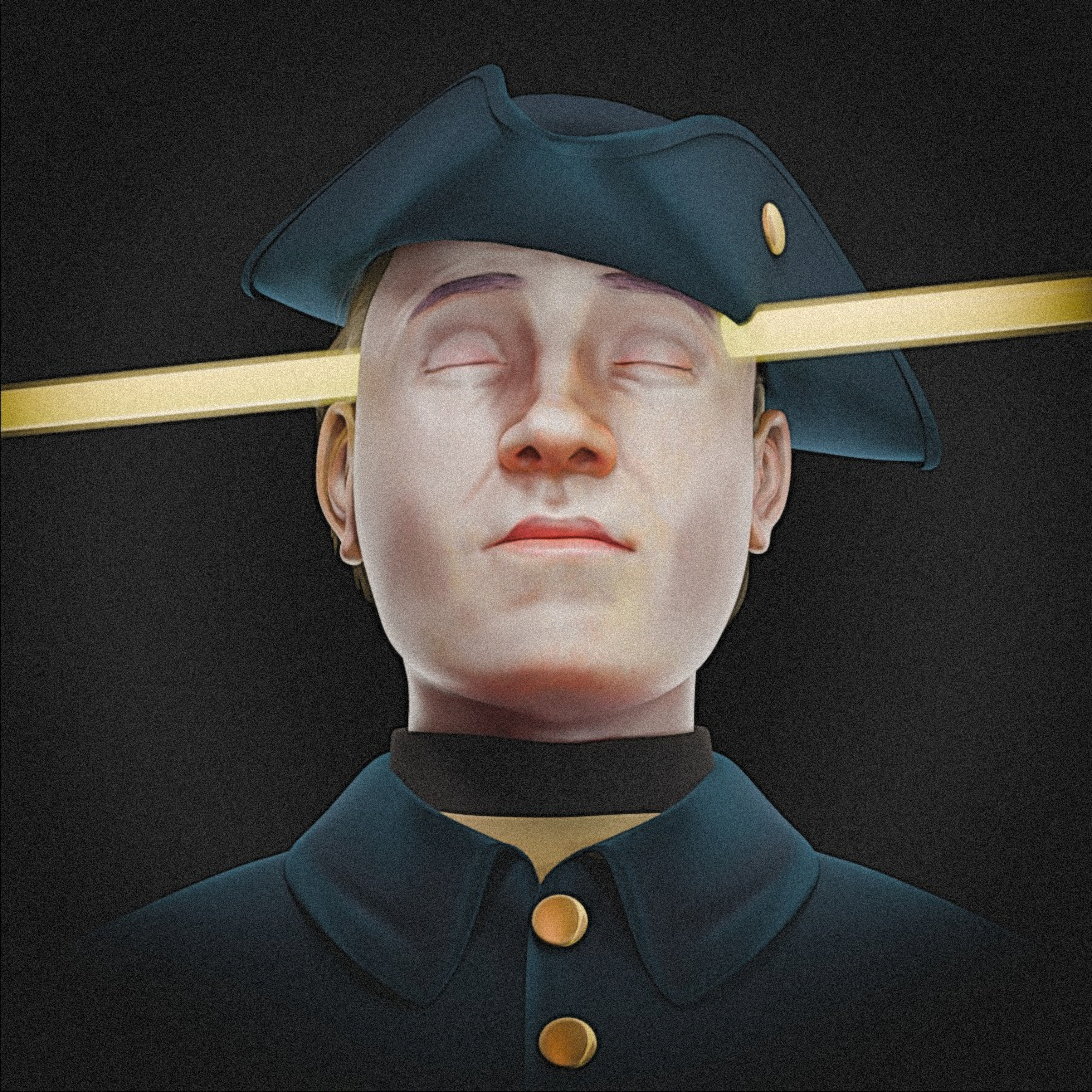
Fig. 51 Frontal and slightly inferior view with full attire and projectile passage.
Although the 1917 exhumation did not attribute significant relevance to the felt hat, explicitly treating it as a secondary element—as indicated by the observation that “nor was there found inside the skull any fragment of the hat that the King supposedly wore at the time of his death” [Key-Aberg_and_Stille_1918_e]—this isolated mention ended up being disproportionately valued by later authors, who began to use it as an evidential anchor. This interpretive shift is problematic, as Key-Åberg’s own report not only avoids employing the hat as a ballistic variable but also bases its conclusions exclusively on the physical marks observed on the skull. Even so, recent works, such as Junno et al. (2022), state that “the projectile that killed Charles also pierced his felt hat leaving a hole of 19 to 19.5 mm in diameter” [Junno_et_al_2022_e], implicitly attributing this measurement to the primary 1917 source, which finds no support in the original text that never associates such a value with the textile artifact. This type of claim suggests a mediated chain of citation, possibly derived from interpretations like that of Nordling (1998), in which the hat had already been unduly elevated to a central element [Nordling_1998_e]. The result is a methodological inversion: an artifact not geometrically validated in relation to the skull begins to define ballistic parameters, while the direct evidence—more robust and explicitly prioritized in the 1917 examination—is relativized or reinterpreted in light of this premise. Furthermore, it remains notable that none of these studies performed the minimum verification necessary to sustain such use of the hat—that is, the spatial and angular correspondence between the hole in the felt and the documented intracranial trajectory—which reinforces the speculative nature of this interpretive line and highlights a persistent methodological gap in the literature.
Precisely considering this gap, the present study positioned the piece as it might have been arranged at the moment of the potential impact (Fig. 51). Although it may appear displaced and excessively close to the eyes, wearing the tricorne hat in this manner finds support in artistic references from the 18th century (https://en.wikipedia.org/wiki/File:Frederic_II_de_prusse.jpg, https://commons.wikimedia.org/wiki/File:Tricorne_18th_century.jpg), 19th century (https://commons.wikimedia.org/wiki/File:Edmund_Fanning_colonial_administrator.jpg, https://commons.wikimedia.org/wiki/File:Fredrik_den_store._Efter_ett_tr%C3%A4snitt_af_B%C3%BCrkner.jpg), and 20th century (https://en.wikipedia.org/wiki/File:US_Army_52416_The_American_Soldier,_1781.jpg).
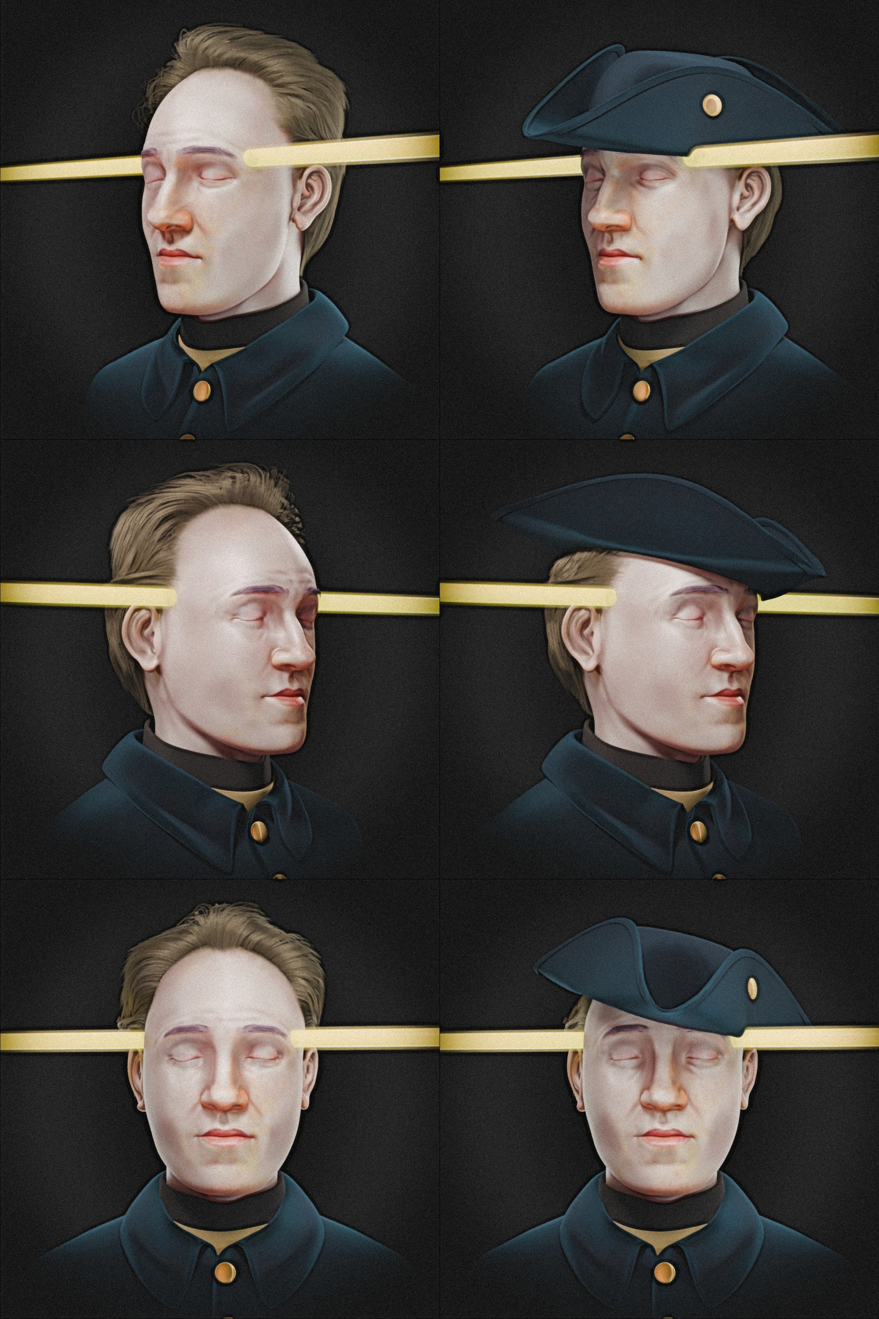
Fig. 52 Images of the bust with the projectile trajectory in versions without and with the felt hat.
A series of images was rendered to illustrate the entry and exit of the projectile on the approximated bust of King Charles XII, serving as a didactic element closely representing what occurred, both in an anatomical context and in relation to the clothing (Fig. 52).
Conclusion
The present study successfully fulfills the objective of performing an unprecedented forensic facial approximation of Charles XII of Sweden, integrating biomechanical and three-dimensional craniometric analyses within an open-source digital environment. Based on the methodology employed, it was possible to reach significant conclusions that enrich the literature and the historical debate surrounding the monarch.
Firstly, the three-dimensional geometric reconstruction of the projectile trajectory robustly corroborated the fundamental observations presented in the historical report of the 1917 exhumation. The digital alignment, based on the center of the wounds identified in the soft tissues, confirmed a practically rectilinear transfixing trajectory, with a nearly horizontal orientation slightly directed toward the rear.
As the main diagnostic novelty, the execution of the USP standard cephalometric analysis revealed, in a quantitative manner, structural characteristics that until now had been approached in a purely descriptive or artistic way. This provided solid craniometric evidence that the King presented a condition of maxillary retrognathism (Class III) associated with mandibular prognathism. Such anatomical findings justify and geometrically validate the facial proportions observed in portraits painted from life and the volumetric characteristics recorded in his death mask, such as the accentuated projection of the labial mass during occlusion.
Furthermore, the work innovates by presenting the first complete three-dimensional anatomical approximation of the skull, brain, and intracranial venous system of Charles XII. This approach allowed not only for the visualization of the monarch’s approximated face with high technical rigor but also for the mapping, with significant precision, of the encephalic and vascular structures severely injured by the impact, such as the frontal and temporal lobes, the corpus callosum, and the middle cerebral artery.
Finally, filling a persistent methodological gap in contemporary literature, the study performed the spatial verification of the royal attire. The precise three-dimensional positioning of the tricorne hat—adjusted in accordance with historical and iconographic references of military use at the time—demonstrated exact geometric compatibility with the projectile’s entry trajectory in the left temporal region.
In summary, the use of the OrtogOnBlender ecosystem and open-source tools proved to be an efficient and highly reproducible methodology for the fields of bioarchaeology and digital forensic science, offering an unprecedented technical assessment that consolidates the integrity of historical data from a strictly geometric and biomechanical perspective.
Data Statement
Acknowledgements
To Dr. Richard Gravalos for providing the tomography of the virtual donor.
References
Baldasso, R. P., Moraes, C., Gallardo, E., Stumvoll, M. B., Crespo, K. C., Strapasson, R. A. P., & de Oliveira, R. N. (2020). 3D forensic facial approximation: Implementation protocol in a forensic activity. In Journal of Forensic Sciences (Vol. 66, Issue 1, pp. 383–388). Wiley. https://doi.org/10.1111/1556-4029.14587
De Greef, S., Claes, P., Vandermeulen, D., Mollemans, W., Suetens, P., & Willems, G. (2006). Large-scale in-vivo Caucasian facial soft tissue thickness database for craniofacial reconstruction. In Forensic Science International (Vol. 159, pp. S126–S146). Elsevier BV. https://doi.org/10.1016/j.forsciint.2006.02.034
Edlow, B. L., Mareyam, A., Horn, A., Polimeni, J. R., Witzel, T., Tisdall, M. D., Augustinack, J., Stockmann, J. P., Diamond, B. R., Stevens, A., Tirrell, L. S., Folkerth, R. D., Wald, L. L., Fischl, B., & van der Kouwe, A. (2019). 7 Tesla MRI of the ex vivo human brain at 100 micron resolution. Cold Spring Harbor Laboratory. https://doi.org/10.1101/649822
Hatton, R.M., Nordlund, S. (2026, March 9). Charles XII. Encyclopedia Britannica. https://www.britannica.com/biography/Charles-XII
Junno, J.-A., Niskanen, M., Maijanen, H., Niinimäki, J., Junno, A., & Oura, P. (2022). The death of King Charles XII of Sweden revisited. PNAS Nexus, 1(5). https://doi.org/10.1093/pnasnexus/pgac234
Key-Åberg, A., & Stille, A. (1918). Konung Karl XII:s banesår: 1917 års undersökning och i samband därmed gjorda iakttagelser. P. A. Norstedt & Söners förlag. Recuperado de https://brf-portalen.se/wp-dragoner/album/k12-banesar/index.html
Kimmerle, E. H., & Baraybar, J. P. (2017). Lesões por Arma de Fogo. In E. H. Kimmerle & J. P. Baraybar, Trauma Esquelético: Identificação de Lesões Ocasionadas por Violação aos Direitos Humanos e Conflitos Armados (pp. 283–464). Editora Unifesp.
Moraes, C. (2023). A Aproximação Facial e a Dinâmica do Acidente de Phineas Gage (1848). figshare. https://doi.org/10.6084/M9.FIGSHARE.24782022
Moraes, C. (2024). A Aproximação Facial da “Vampira” de Veneza (Séc. XVI-XVII). figshare. https://doi.org/10.6084/M9.FIGSHARE.25447270
Moraes, C., Bezzi, L., & Bezzi, A. (2024). A Aproximação Facial e a Análise Estrutural do Crânio do Homem de Piltdown - Fraude (1912). figshare. https://doi.org/10.6084/M9.FIGSHARE.24937035
Moraes, C., & Habicht, M. (2024). A Aproximação Facial Forense de Seqenenre-Taa-II (c. 1558-1553 a.C). figshare. https://doi.org/10.6084/M9.FIGSHARE.25945906
Moraes, C., & Moura, L. (2025). 3D Digital Analysis of the Anatoli Bugorski Case (1978). figshare. https://doi.org/10.6084/M9.FIGSHARE.29973373
Moraes, C., & Suharschi, I. (2022). Mensuração de Dados Faciais Ortográficos em Moldavos e Comparação com Outras Populações. figshare. https://doi.org/10.6084/M9.FIGSHARE.20089754. https://ortogonline.com/doc/pt_br/OrtogOnLineMag/4/Moldavos.html
Moraes, C., Beaini, T. L., Steffensen, T. H., & Dalstra, M. (2022). A Aproximação Facial de uma Vítima da Batalha de Gotland (1361). Figshare. https://doi.org/10.6084/M9.FIGSHARE.21432384
Moraes, C., Habicht, M. E., Galassi, F. M., Varotto, E., & Beaini, T. (2023). Pharaoh Tutankhamun: a novel 3D digital facial approximation. Italian Journal of Anatomy and Embryology, 127(1), 13–22. https://doi.org/10.36253/ijae-14514
Moraes, C., Habicht, M. E., Artico, M., Forte, F., Varotto, E., & Galassi, F. M. (2024). The mummy of Pharaoh Amenhotep III (reigned ca. 1388–1351 BC) and its facial approximation: An anatomical approach. Clinical Anatomy, 38(2), 211–215. https://doi.org/10.1002/ca.24251
Moraes, C., Moura, L., Beaini, T. L., & Paiva Curi, J. (2025). A Aproximação Facial do Homem de Porsmose (ca. 5500 AP) [Dataset]. figshare. https://doi.org/10.6084/M9.FIGSHARE.28207625
Nasca, G., Ioime, G., Pellegrini, A., Ianora, A. A. S., Introna, F., Di Gioia, E., & Galantucci, L. M. (2026). A novel forensic protocol for age reversal facial approximation from elderly skulls using computed tomography scans, CAD techniques and anthropometric data. Forensic Science International, 385, 112960. https://doi.org/10.1016/j.forsciint.2026.112960
Nordling, C. O. (1998). The death of King Charles XII — The forensic verdict. Forensic Science International, 96(2–3), 75–89. https://doi.org/10.1016/s0379-0738(98)00110-8
Tank, P. W., & Gest, T. R. (2009). Atlas de anatomia humana (A. L. Werneck & H. Werneck, Trads.; 1ª ed.). Artmed.
Universidade de São Paulo. (2021). Análise cefalométrica simplificada. e-Disciplinas USP. https://web.archive.org/web/20230610023749/edisciplinas.usp.br/pluginfile.php/5214352/mod_resource/content/2/Estudo%204.pdf