Rest in Face: The Forensic Approximation of Individual 157 from the Račeša Necropolis (13th–14th Century)
Abstract
The Račeša necropolis in Croatia is a complex archaeological site dated from the 13th to 16th centuries, initially associated with the Templars and later with the Order of the Knights Hospitaller of Saint John of Jerusalem. Excavations between 2012 and 2023 revealed an extensive architectural structure and 181 inhumations, most following the traditional Christian rite. However, grave 157 stood out for its atypical nature: a middle-aged man (40-50 years), stature of 164 cm, with evidence of occupational stress and intense physical violence, including old lesions on the maxilla and ribs, and perimortem traumas on the cranium caused by a sharp object. The burial deviated from the standard, with the thorax in a prone position (face down) and the cranium removed and repositioned 30 cm from the neck, suggesting postmortem manipulation motivated by beliefs in revenants (vampires) according to Slavic traditions. Given this singular profile, this study applied forensic facial approximation to the fragmented cranium, reconstructed via computed tomography and open-source digital tools such as OrtogOnBlender. The cranium, segmented into 21 parts, underwent anatomical repositioning, with soft tissue projections based on European population averages, soft tissue thickness markers, and anatomical deformation using a virtual donor. Three images were generated: two objective ones in grayscale without facial hair and one artistic with speculative coloration. Additionally, the brain volume of 1488 cm³ indicated mild megalencephaly (+2.59 standard deviations relative to the modern male average).
Keywords: Forensic facial approximation, free software, open source, anthropology, vampire.
Important
This material uses the following Creative Commons license: Attribution 4.0 International (CC BY 4.0).
Attention
Should you encounter any errors in the text, kindly report them to the author; he may be contacted through the academic social media platforms mentioned at the beginning of the chapter.
Introduction
The necropolis of Račeša, Croatia, is a complex archaeological site whose excavations have revealed a remarkable occupational sequence. Initially associated with a Templar property, and subsequently under the dominion of the Order of the Knights Hospitaller of Saint John of Jerusalem, the site has an established chronology between the 13th and 16th centuries. The research, which took place between 2012 and 2023, brought to light an extensive architectural complex and 181 inhumations, the majority following the traditional Christian rite, positioned in a west-east direction [Sarkic_et_al_2024_b].
However, one finding in particular deviated drastically from the standard: grave number 157. This grave was discovered in a layer beneath the church floor, positioned in a less favored spot next to the southern wall of the nave. Osteological analysis of the skeleton revealed it to be a middle-aged man, with an estimated age between 40 and 50 years. His stature was calculated at 164.03 cm. The individual’s biological profile is marked by prominent markers of occupational stress and a history of intense physical violence.
The evidence of trauma is multiple and dates from different moments in his life, including an old sharp-force injury on the maxillary bone and rib fractures that were still in the process of healing at the time of death. The fatal injury, however, resulted from acute interpersonal violence, attested by two perimortem traumatic lesions on the back of the skull, caused by a sharp object, with no signs of healing. In addition to the violent death, the old injuries suggest that this man may have lived with a partially disfigured face.
The positioning of the body in the tomb is the most intriguing characteristic. Grave 157 presented an atypical burial, notably by the inversion of the thorax to a prone position (face-down) and the removal and repositioning of the skull approximately 30 cm from the neck. The absence of cut marks suggests that this postmortem manipulation occurred early on, while the soft tissues were already putrefying, but before the complete degradation of the ligaments. Such deviant burial practices, associated with individuals with violent deaths or a history of conflict, suggest an intervention motivated by beliefs in revenants (vampires), according to Slavic tradition.
Given the singular profile of the individual from grave 157—a man marked by violence in life and manipulated in death—it becomes imperative to reestablish his visual identity. The objective of this work is, therefore, to determine the probable facial appearance of this individual, providing a new perspective for the understanding of his potential social exclusion and the community’s reaction to his death. To this end, the team reconvened to apply the forensic facial approximation technique to the skull, transforming the skeletal data into a face that transcends the archaeological record.
Materials and Methods
The skull from tomb 157 was found fragmented [Sarkic_et_al_2024_b] and subsequently manually reconstructed by specialists, undergoing a computed tomography scan that digitized it in 3D. A video in RGB color format (8 bits) served as the basis for the three-dimensional recovery of the structure. Initially, the file in Audio Video Interleave (AVI) format, compressed via the Microsoft Video 1 codec and with dimensions of 1040 x 1532, was converted into a sequence of grayscale images and, subsequently, into a DICOM sequence (voxel dimensions: 0.23364, 0.23364, 0.6), using specific tools for this purpose [Moraes_et_al_2021_b] from the OrtogOnBlender add-on (http://www.ciceromoraes.com.br/doc/pt_br/OrtogOnBlender/index.html).
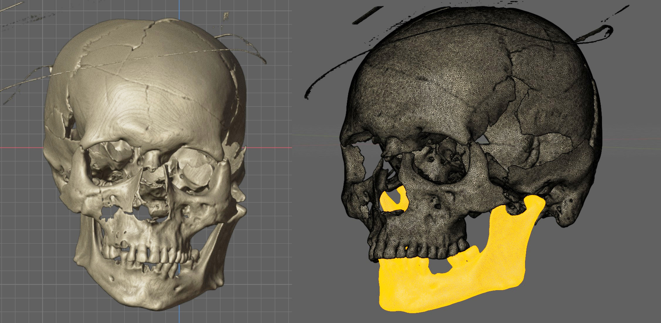
Fig. 8 On the left, the DICOM file reconstructed in 3D. On the right, the mesh being edited for mandible segmentation.
Once converted to DICOM, it was possible to reconstruct the 3D mesh using a segmentation threshold (Hounsfield-equivalent factor) of 53 [Moraes_et_al_2021b_b], due to the RGB origin of the material (Fig. 8, left). The resulting 3D mesh enabled the segmentation of the anatomical pieces for the subsequent adjustments that were performed thereafter (Fig. 8, right).
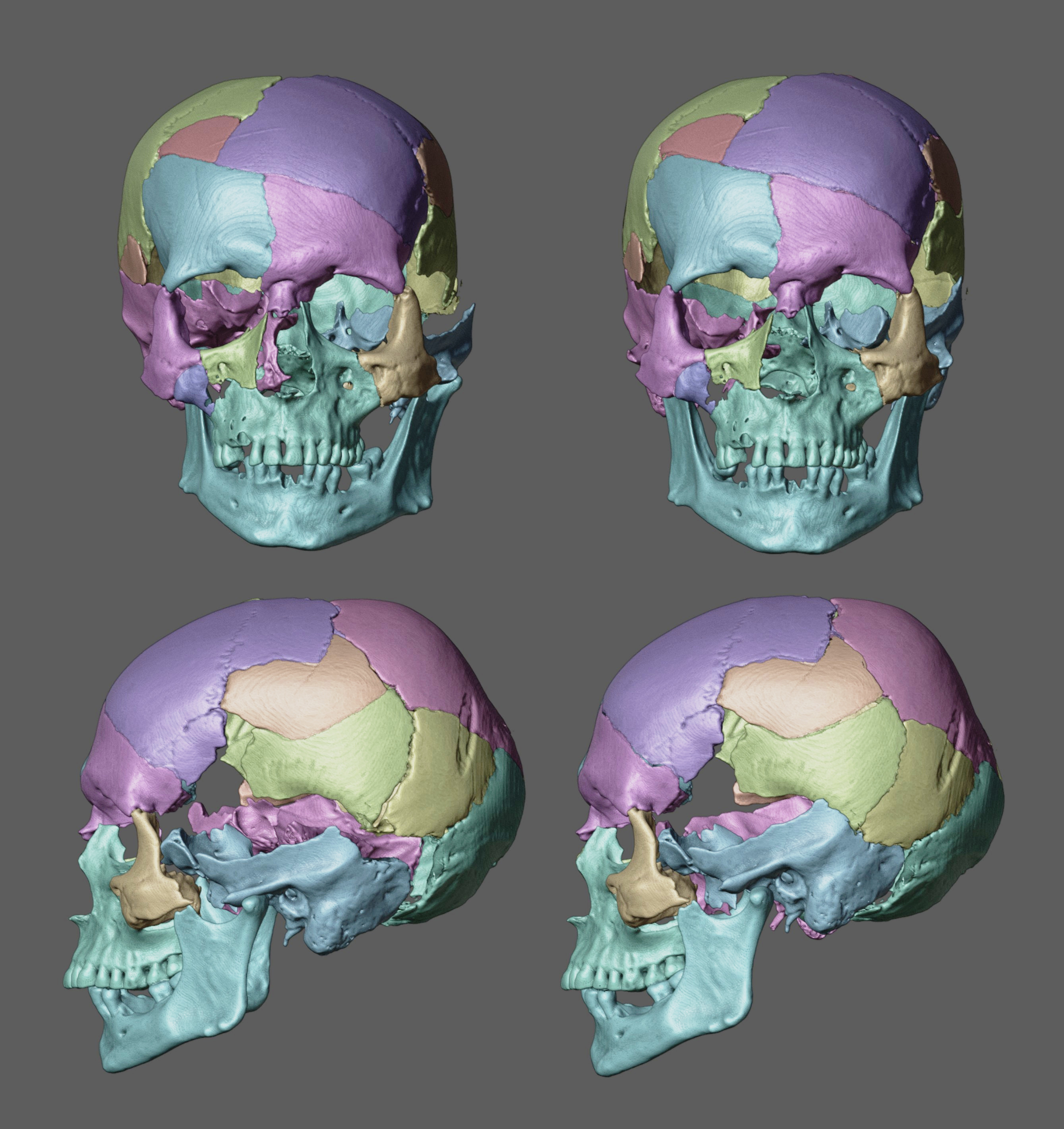
Fig. 9 The left side (top and bottom) presents the original 3D reconstructed mesh segmented. The right side (top and bottom) shows the reassembled segments with anatomical coherence.
The skull was segmented into 21 parts (Fig. 9, left) and repositioned according to criteria of structural and anatomical coherence (Fig. 9, right). A comparative animation showing two states — the original fragmented condition and the reorganized one — facilitated the dynamic observation of the fragment positioning. This analysis, combined with the operator’s experience and structural comparison with a reference skull (virtual donor), enabled the definitive fixation of the cranial configuration that served as the basis for the subsequent facial approximation.
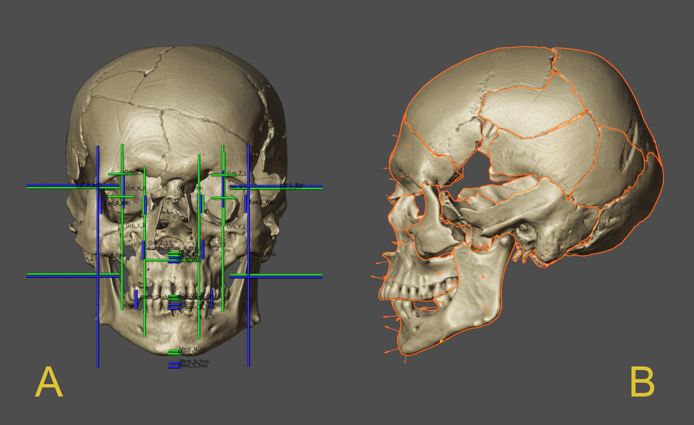
Fig. 10 A) Projections based on average anatomy. B) Soft-tissue depth markers.
Once the definitive cranial configuration was established, anatomical landmarks were placed to allow anthropometrically-based projections to indicate the expected averages and proportions for both the existing structures and those to be reconstructed (video tutorials available here: class 1, class 2). The resulting guidelines delineate the anticipated anatomical spaces, indicating, for example, the position of the eyeballs, the limits of the nasal wings and nasal base, the horizontal boundaries of the lips, and others. Additionally, these projections also reveal the expected dimensions for present bony structures, such as the mandibular contours [Moraes_and_Suharschi_2022_b].
In the present case, the mandible slightly exceeded the average value along the X-axis at the gonion region, remained within the expected average for height, and displayed a proportional dimension (in blue) below the reference mean (in green). This configuration is explained by the frontomalare orbitale measurement of 101.5 mm, which is above the general average of 96.6 mm (https://bit.ly/3NRw2KW), indicating that the skull is slightly larger than average in the transverse axis (X) while preserving the expected dimensions in the vertical axis (Y) (Fig. 10, A).
For the projection of facial boundaries, soft-tissue depth markers derived from population averages of living European males aged 40–49 years [De_Greef_et_al_2006_b] were distributed across the cranial surface. These data are consistent with the forensic anthropological findings reported by [Sarkic_et_al_2024_b] (Fig. 10, B).
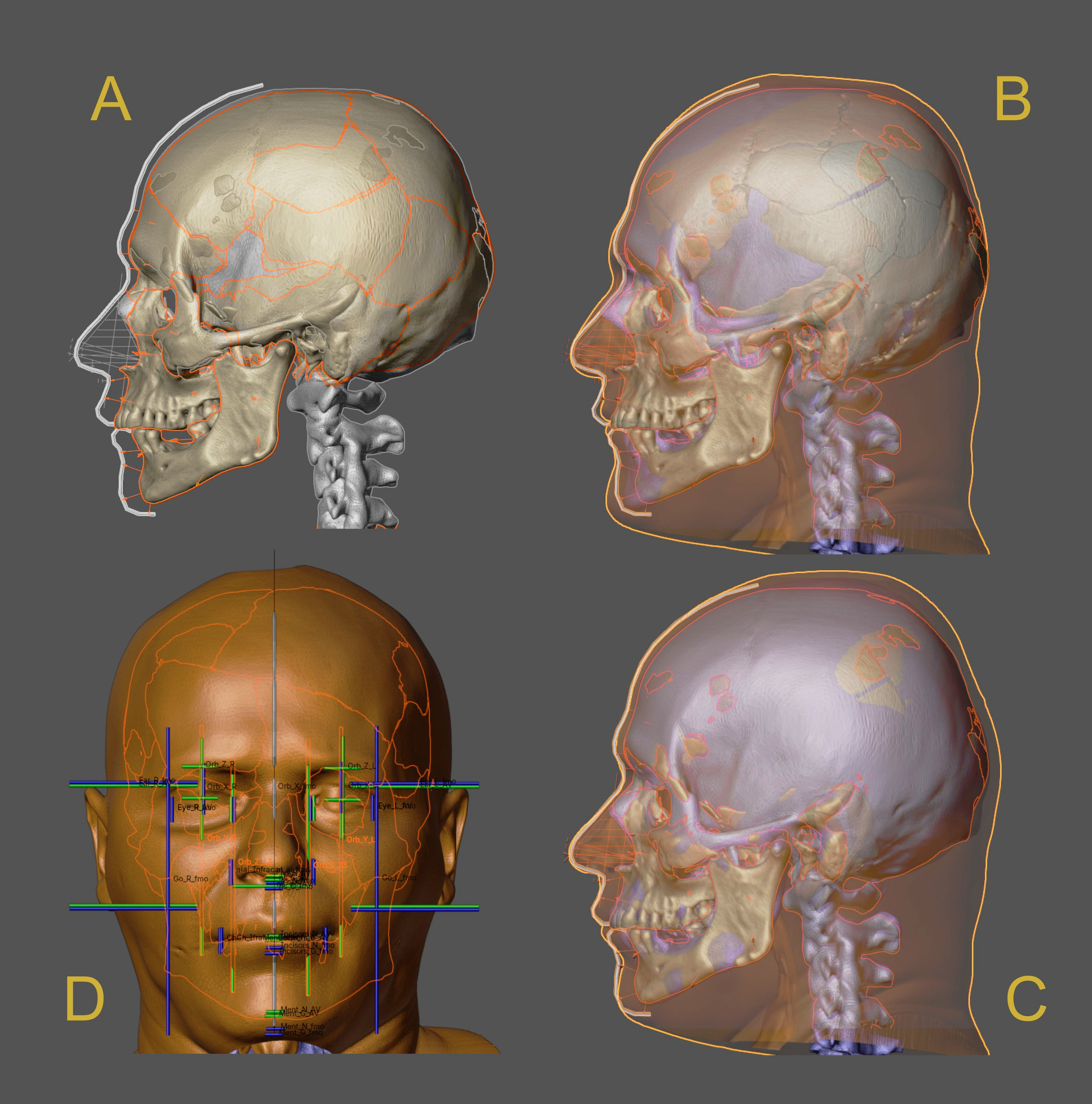
Fig. 11 A) Soft-tissue depth markers, nasal projection, and facial profile tracing. B) Original composite head of the virtual donor. C) Composite head adjusted to the skull of individual 157 – Anatomical deformation. D) Frontal view of the anatomical deformation with limit lines, demonstrating structural coherence between the two sources.
Although reassembled and anatomically coherent, the skull of individual 157 exhibited some structural absences that were filled using the skull of a virtual donor, which then enabled nasal projection according to data obtained from computed tomography scans of living individuals [Moraes_and_Suharschi_2022_b]. The soft-tissue depth markers, together with the nasal projection data, allowed the tracing of the facial profile (Fig. 11, A).
To further supplement the facial projection data, the anatomical deformation approach was employed [Quatrehomme_et_al_1997_b], which involves importing a complete head from a virtual donor—composed of meshes of soft tissue, cranium, and endocranium—and adapting it until the donor’s skull matches that of individual 157, thereby reflecting the corresponding adjustment in the soft tissue and, consequently, in the face (Fig. 11, B and C).
Laterally, the results of the anatomical deformation proved compatible with the previously established profile projection (Fig. 11, C), and this coherence also extended to the frontal view of the face in relation to the limit lines based on soft-tissue measurements, correctly corresponding to the mid-orbital points, the nasal limits, the lip boundaries, and the ear positions (Fig. 11, D).
Tip
For replication purposes, classes on nasal projection (https://youtu.be/F205kLQ–Oo) and anatomical deformation (https://youtu.be/xig5_EcIFWA).
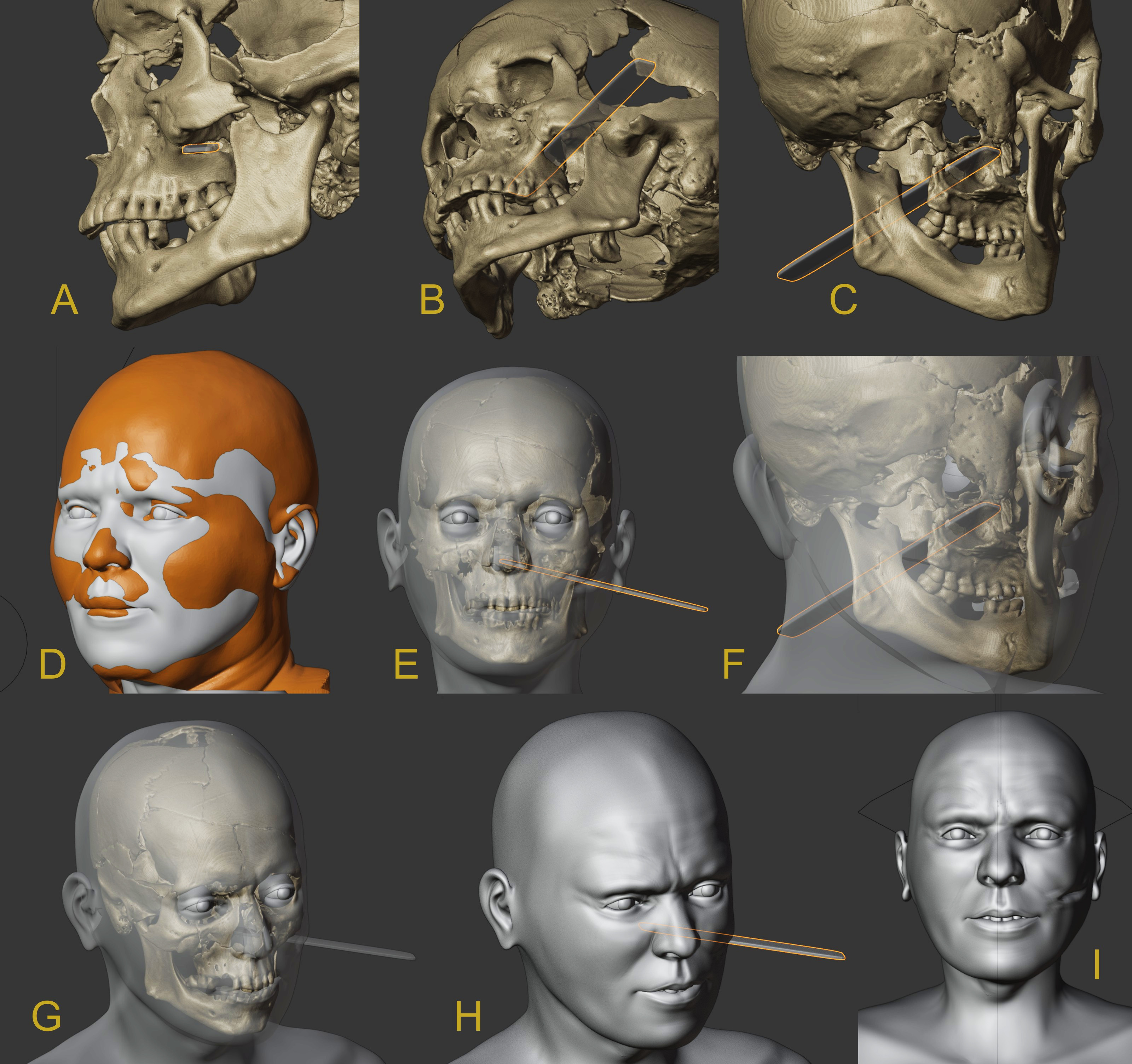
Fig. 12 A, B, C) Structure of the penetrating-cutting trajectory. D) Anatomical deformation/projections with the final basic face (in gray). E, F, G) Visualization of the transparent face with the volumetric trajectory of the lesion. H, I) Face with digital sculpture details and scar in the lesion region.
A structure was positioned in the lesion caused by, probably, a penetrating-cutting object that affected the zygomatic process from the external aspect, penetrating through it and exiting slightly above the palatine process of the maxilla (Fig. 12, A, B, C). Following the creation of the final basic face (Fig. 12, D), it was possible to identify the location of the lesion in the soft tissue (Fig. 12, E, F, G) and, from there, finalize the details of the facial sculpture with the resulting scar (Fig. 12, H, I).
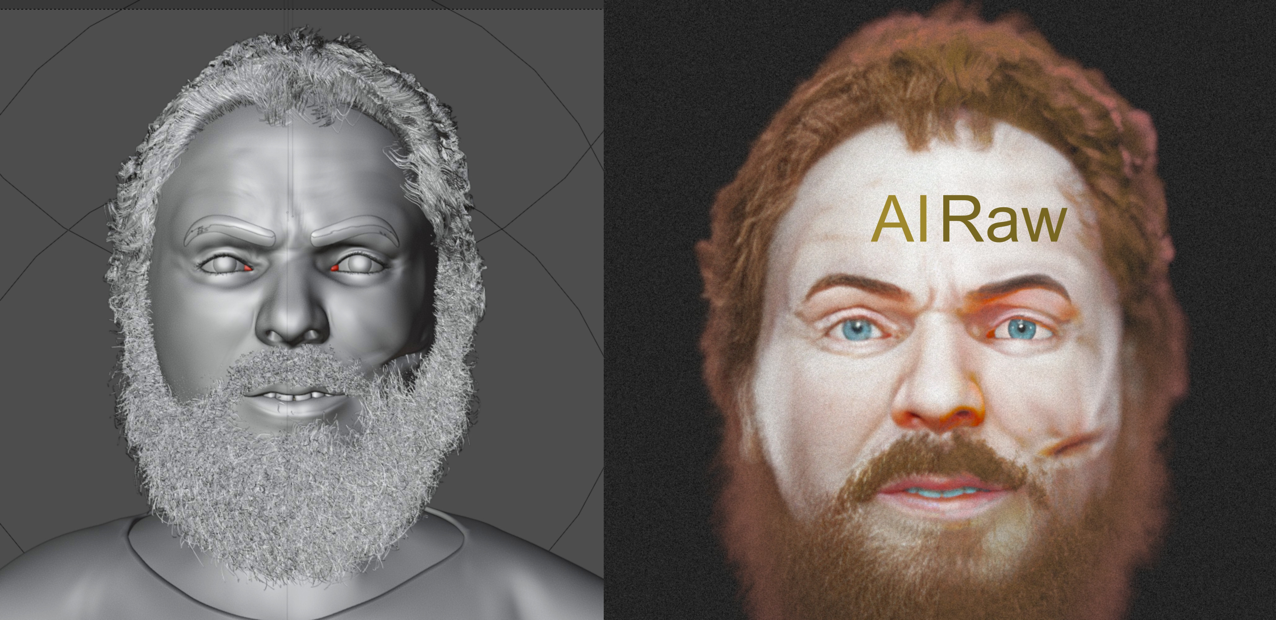
Fig. 13 On the right) Solid composition of the face with facial hair and attire. On the left) Comparison between the original version (Raw) and adjustments controlled by artificial intelligence (AI).
Subsequently, details related to more speculative elements were configured, such as facial hair and attire (Fig. 13, on the left). To finalize, the scene was illuminated, colors adjusted, opting for eyes with gray-bluish coloration and reddish-brown hair. Finally, small details were adjusted using artificial intelligence through the Codeformer library [Zhou_et_al_2022_b], directly via OrtogOnBlender XP, in an assisted manner and using a weight of 0.99, that is, very close to the original image (1.00), which can be perceived by the small variation between the original (Raw) and the adjusted version (AI) (Fig. 13, on the right).
Results and Discussion
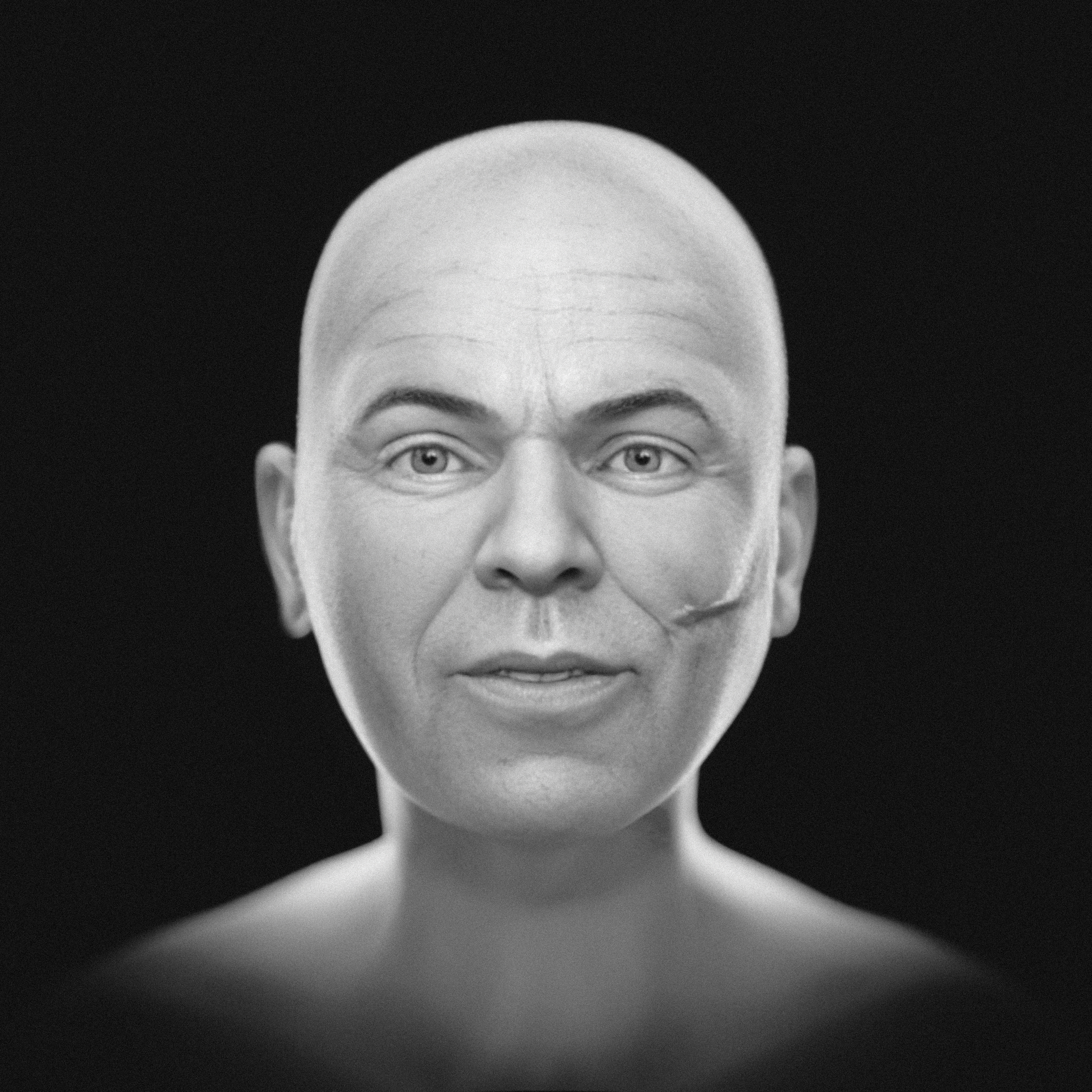
Fig. 14 Grayscale face without hair and beard - frontal.
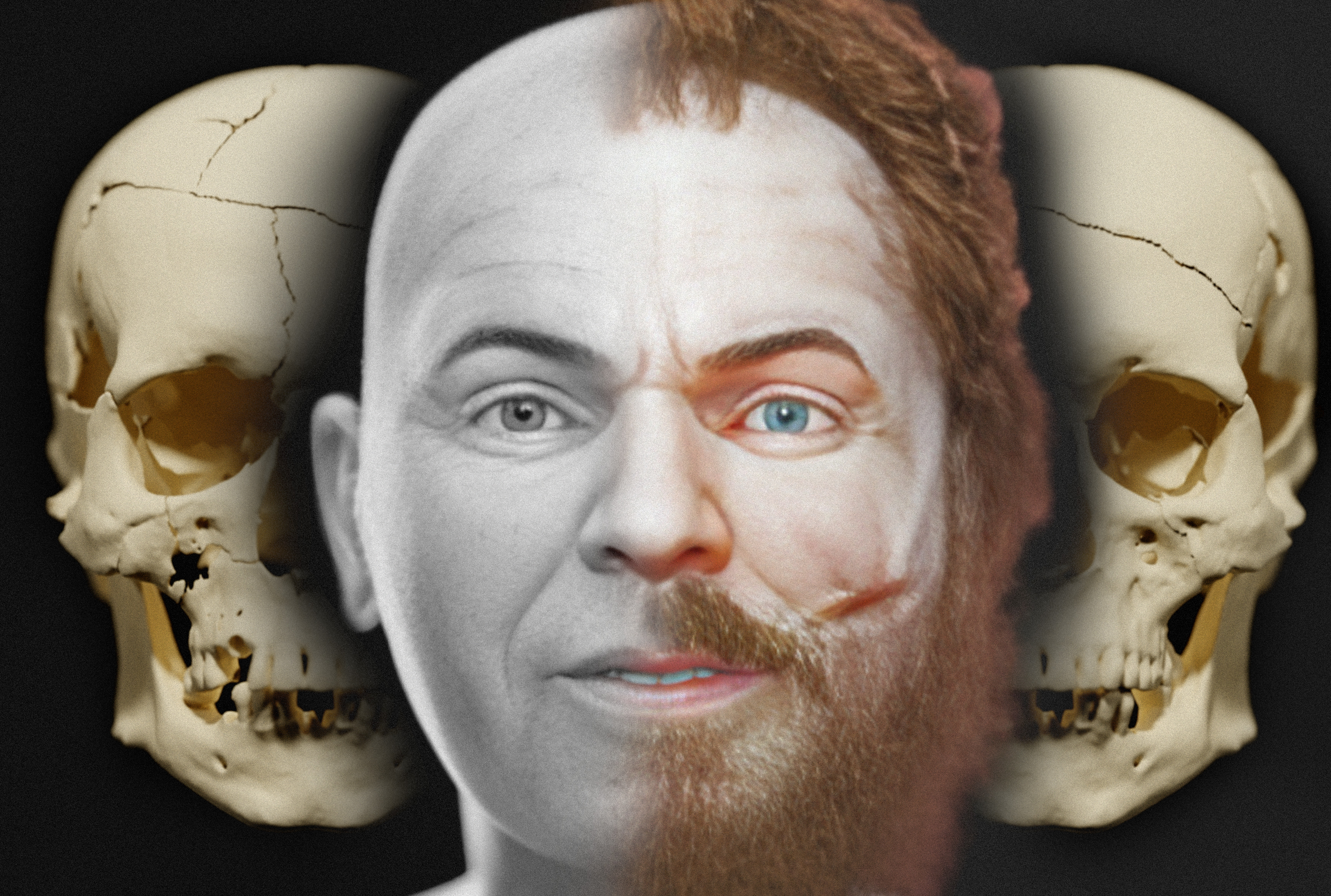
Three images were generated at the end of the process, two related to a more objective version, in grayscale and without facial hair (Fig. 14, Fig. 15) and one with more artistic and therefore speculative elements, with coloration of the skin, eyes, and hair (Fig. 16).
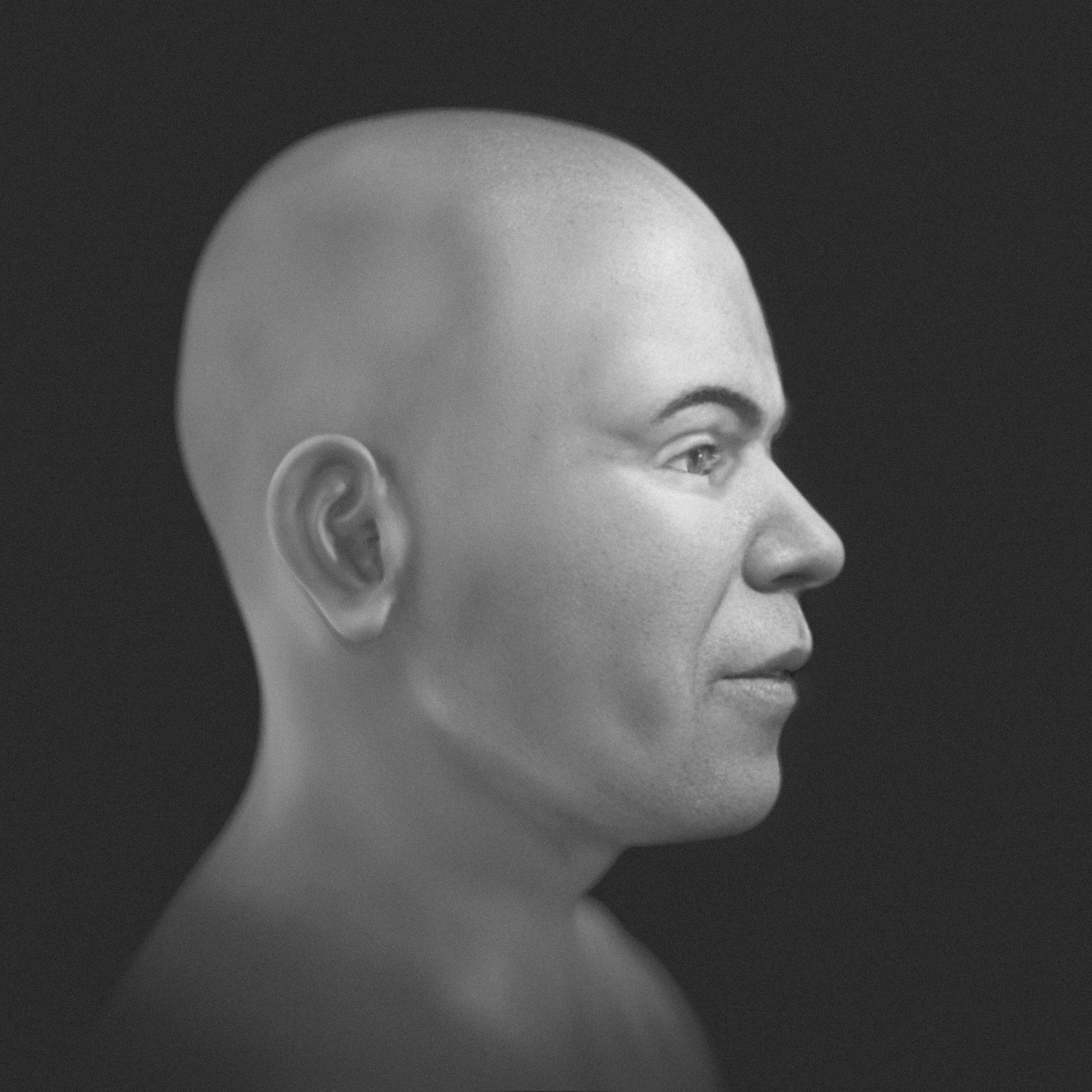
Fig. 15 Grayscale face without hair and beard - profile.
It should be noted that forensic facial approximation contains some limitations related to the precision of fine details, such as expression marks, eye shape, etc. However, regarding the position of the eyes, lip dimensions, and nose shape, in general terms, the structures are significantly compatible with the individual in life, to the point of providing data for the recognition of crime victims and subsequent identification, as illustrated in a forensic case involving one of the authors (C.M.) [Baldasso_et_al_2020_b].
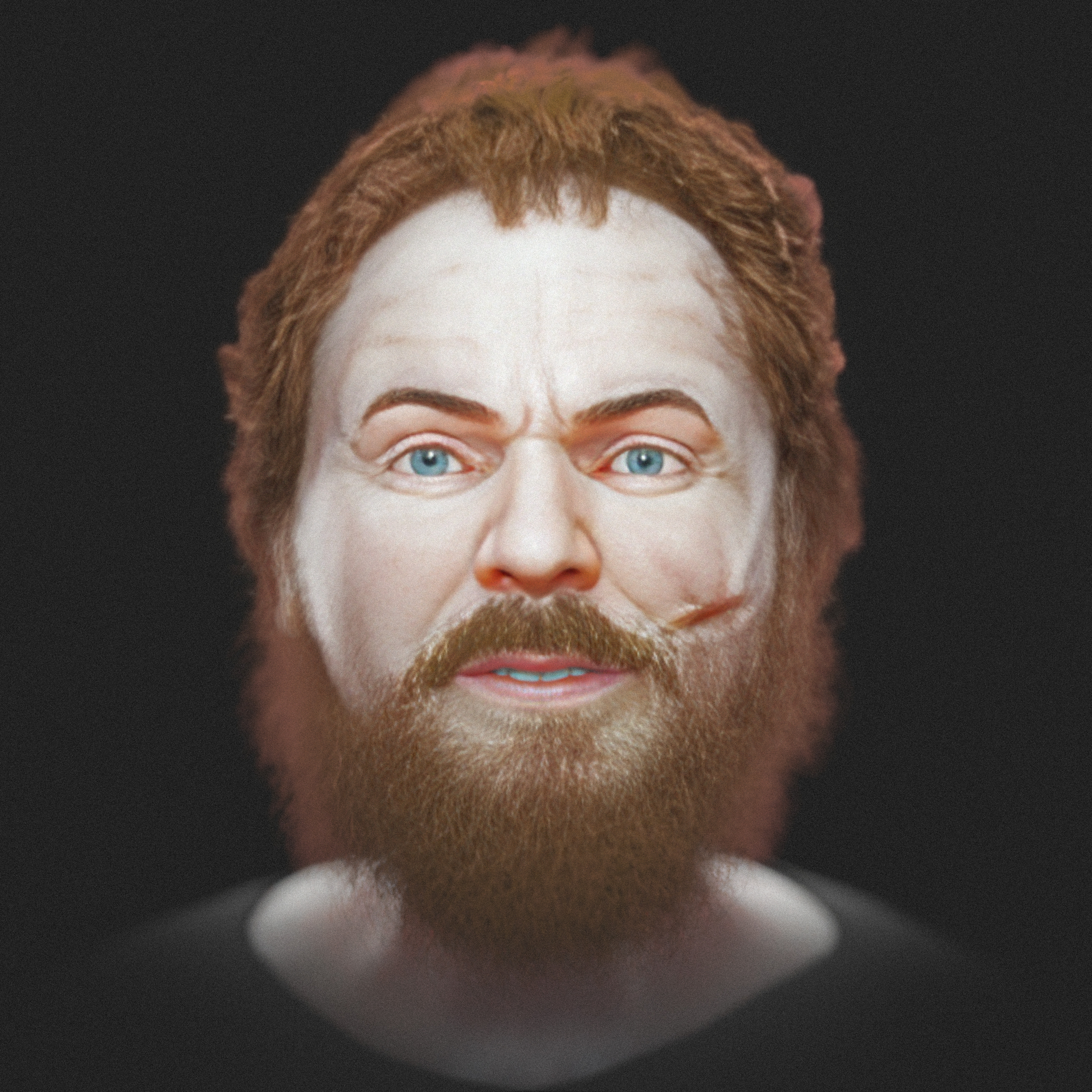
Fig. 16 Colored version with more subjective/artistic elements.
In addition to the facial approximation, the endocranium of Individual 157 was also segmented, resulting in ~1650 cm³. When converted to brain volume (-9.81%) [Moraes_et_al_2023_b], it resulted in 1488 cm³, which compared to the modern male average of 1234 cm³ (± 98) [Ritchie_et_al_2018_b] is at +2.59 standard deviations, indicating mild megalencephaly. This finding is reported as a morphological observation and should not be interpreted as indicative of cognitive or behavioral traits.
Conclusion
This study demonstrates the effectiveness of forensic facial approximation in reconstructing the visual identity of archaeological individuals, such as Individual 157 from the Račeša necropolis, marked by violence in life and postmortem manipulation. The integration of digital techniques, such as 3D segmentation and anatomical deformation, allowed for the generation of facial representations compatible with anthropological data, highlighting lesions and scars that suggest a conflictual existence and possible social stigmatization. Despite the inherent limitations in the precision of speculative details, the results provide valuable insights into deviant funerary practices linked to beliefs in revenants, enriching the historical and cultural understanding of the Croatian medieval period.
Acknowledgements
To Dr. Richard Gravalos for providing the tomography of the virtual donor. To Michael Havis for proposing the idea of the project and putting the authors in contact.
References
Baldasso, R. P., Moraes, C., Gallardo, E., Stumvoll, M. B., Crespo, K. C., Strapasson, R. A. P., & de Oliveira, R. N. (2020). 3D forensic facial approximation: Implementation protocol in a forensic activity. In Journal of Forensic Sciences (Vol. 66, Issue 1, pp. 383–388). Wiley. https://doi.org/10.1111/1556-4029.14587
De Greef, S., Claes, P., Vandermeulen, D., Mollemans, W., Suetens, P., & Willems, G. (2006). Large-scale in-vivo Caucasian facial soft tissue thickness database for craniofacial reconstruction. In Forensic Science International (Vol. 159, pp. S126–S146). Elsevier BV. https://doi.org/10.1016/j.forsciint.2006.02.034
Quatrehomme G, Cotin S, Subsol G, Delingette H, Garidel Y, Grévin G, Fidrich M, Bailet P, Ollier A. A fully three-dimensional method for facial reconstruction based on deformable models. J Forensic Sci. 1997 Jul;42(4):649-52. PMID: 9243826.
Moraes, C., Dallazen, E., Dornelles, R., & Da Rosa, E. (2021). Conversão de Imagens em Arquivos DICOM com o OrtogOnBlender. figshare. https://doi.org/10.6084/M9.FIGSHARE.14445366. https://ortogonline.com/doc/pt_br/OrtogOnLineMag/3/ImagemDICOM.html
Moraes, C., Dornelles, R., & Rosa, E. D. (2021). Sistema de Reconstrução de Tomografia Computadorizada Baseado no Slicer 3D e no DicomToMesh. figshare. https://doi.org/10.6084/M9.FIGSHARE.13513890. https://ortogonline.com/doc/pt_br/OrtogOnLineMag/2/Slicer.html
Moraes , C., Habicht , M. E., Galassi, F. M., Varotto, E., & Beaini , T. (2023). Pharaoh Tutankhamun: a novel 3D digital facial approximation. Italian Journal of Anatomy and Embryology, 127(1), 13–22. https://doi.org/10.36253/ijae-14514
Moraes, C., & Suharschi, I. (2022). Mensuração de Dados Faciais Ortográficos em Moldavos e Comparação com Outras Populações. figshare. https://doi.org/10.6084/M9.FIGSHARE.20089754. https://ortogonline.com/doc/pt_br/OrtogOnLineMag/4/Moldavos.html
Ritchie, S. J., Cox, S. R., Shen, X., Lombardo, M. V., Reus, L. M., Alloza, C., Harris, M. A., Alderson, H. L., Hunter, S., Neilson, E., Liewald, D. C. M., Auyeung, B., Whalley, H. C., Lawrie, S. M., Gale, C. R., Bastin, M. E., McIntosh, A. M., & Deary, I. J. (2018). Sex Differences in the Adult Human Brain: Evidence from 5216 UK Biobank Participants. In Cerebral Cortex (Vol. 28, Issue 8, pp. 2959–2975). Oxford University Press (OUP). https://doi.org/10.1093/cercor/bhy109
Šarkić, N., Cighetti, R., Schendzielorz, S.-K., & Mihaljević, M. (2024). REST IN PIECES: AN ATYPICAL BURIAL FROM THE NECROPOLIS IN RAČEŠA (CROATIA), 13TH–16TH CENTURY. In J. Belaj, Z. Hunyadi, T. Tkalčec, S. Krznar, T. S. Ivančan, T. Karavidović, T. Kokotović, & S. Stingl (Eds.), MILITARY ORDERS AND THEIR HERITAGE (pp. 233–244). Institute of Archaeology.
Zhou, S., Chan, K. C. K., Li, C., & Loy, C. C. (2022). Towards Robust Blind Face Restoration with Codebook Lookup Transformer (Version 2). arXiv. https://doi.org/10.48550/ARXIV.2206.11253