Forensic Facial Approximation of Kennewick Man (~8,370 BP)
Abstract
This study presents a digital forensic facial approximation of the individual known as Kennewick Man (~8,370 BP), using exclusively publicly available data for didactic and methodological purposes. The reconstruction was carried out from three-dimensional models of the skull and mandible, scaled according to published osteometric measurements and processed in a free and open-source software environment using the OrtogOnBlender add-on for Blender. The method integrates craniofacial anatomical projections, soft tissue depth markers derived from modern Asian samples, nasal projection estimates, and anatomical deformation of a virtual donor based on computed tomography data. Three sets of results were produced: a statistically grounded basic facial structure, a grayscale forensic approximation following classical forensic standards, and a colored artistic version, the latter explicitly identified as speculative. Additionally, endocranial volume was estimated from the digital endocast, yielding values consistent with modern male variation. The study demonstrates the feasibility of producing reproducible, transparent, and educational forensic facial approximations even in the absence of direct access to physical human remains.
Keywords: Forensic facial approximation, Kennewick Man, 3D facial reconstruction, Digital forensic anthropology, Open-source software.
Warning
This work is independent and has no affiliation with the institution that studied the remains of Kennewick Man. The motivating factor of this chapter is the creation of educational material for teaching facial approximation techniques, by testing the possibility of reconstructing a face using data originally available online, ranging from 3D scans to academic articles.
Introduction
A nearly complete skeleton, composed of 350 fragments representing 143 elements [Chatters_2017_a], was discovered on the banks of the Columbia River in Kennewick, Washington state (USA), in July 1996 [Chatters_2000_a], by two university students who were walking in the area. They called the police, who, given the characteristics of the find, contacted the local archaeologist, Dr. James Chatters [Preston_2014_a].
They did not know it, but they were facing what the Smithsonian considered the most important human skeleton ever found in North America (Preston, 2014).
Over many years of studies and legal twists and turns—which even involved the FBI—it became possible to determine various characteristics of the bones. It was a male individual, aged between 35 and 45 years, with a height of 173.1 cm (± 3.4 cm), an estimated weight of 70 to 75 kg, and moderate robusticity [Chatters_2000_a]. He was right-handed and died around 8,368 ± 21 BP [Chatters_2017_a].
In the initial analyses, Chatters observed that, although the bone structure indicated an Asian or Pacific coast origin, it differed from modern United States Indigenous peoples, presenting features compatible with Polynesian populations, particularly Eastern Pacific islanders and Jōmon/Ainu peoples [Chatters_2000_a]. These observations were complemented by a 2015 mitochondrial DNA study, which confirmed the Asian origin and indicated statistical proximity to the Colville people and individual proximity to the Anzick-1 sample [Rasmussen_et_al_2015_a].
Regarding diet, isotopic analyses of the bones revealed that the diet of the Kennewick Man, as he became known, was predominantly based on marine-derived proteins [Chatters_2000_a], in addition to consumption of potable water from glacial sources [Chatters_2017_a]. He presented minor anomalies in the feet, mild osteoarthritis in the knee and right shoulder joints, as well as evidence of injuries sustained throughout life, such as a small cranial depression, rib fractures, a fracture in the right scapula, and most notably, a spear projectile wound with part of the point still lodged in the right ilium. According to the research, there is consensus that the fractures in the ribs and ilium occurred in early adulthood, while the injuries to the skull and shoulder were close to the time of death (Chatters, 2017). Severe exostosis was also observed in the bones of the external auditory canal, with both nearly obstructed [Chatters_2000_a], a condition compatible with the known “surfer’s ear,” typically caused by frequent immersion in cold water [Preston_2014_a].
Since its discovery, the Kennewick remains became involved in intense legal disputes, particularly related to the Native American Graves Protection and Repatriation Act (NAGPRA), which sought the repatriation of the mortal remains. The case mobilized 93 lawyers and included an FBI investigation involving Drs. James Chatters and Floyd Johnson, in the initial context of forensic osteology. In 2002, the court ruled that the bones did not belong to any modern tribe, rendering NAGPRA inapplicable. An appeal attempt was rejected in 2004, consolidating the scientists’ victory. Following the final verdict, an extensive analysis project was carried out, including detailed documentation, external and internal 3D scanning via computed tomography, 3D printing of the pieces (including the projectile in the ilium), and a forensic facial reconstruction. Initially modeled in clay by a technical team, the face was later refined by StudioEIS, which aged the features, modeled hair and beard, and applied pigmentation for museum exhibition. The chosen appearance was compatible with the Jōmon people, who emerged in Japan approximately 12,000 years ago, based on photographic documentation and possible links to the Ainu—consistent with the craniometric analyses [Preston_2014_a].
Important
An initial facial approximation had been created for the documentary “Mystery of the First Americans,” carried out by Thomas McClelland under the guidance of Dr. James Chatters. This version featured a monochromatic head with a significantly pronounced nose and was presented as early as 2000 [Chatters_2000b_a]. In contrast, the second approximation, mentioned earlier, displayed a less pronounced nose and was presented in 2014 [Preston_2014_a].
However, the 2015 genetic study changed the scenario: by confirming affinity with modern Native American populations, it prompted political action that reclassified the remains as Native American, resulting in the application of NAGPRA and the repatriation of the skeleton. The reburial took place on February 18, 2017, at an undisclosed location [Mabbutt_2017_a].
Materials and Methods
The work presented in this manuscript was conducted using open-source software. The primary tool employed is OrtogOnBlender (https://www.ciceromoraes.com.br/doc/pt_br/OrtogOnBlender/index.html), an add-on for Blender that enables this software to acquire capabilities absent in the original program, such as reconstructing computed tomography scans, performing photogrammetry, utilizing forensic facial reconstruction tools, and others, including enhancing facial details through artificial intelligence. Although this work was executed on Linux Ubuntu 24.04, since all the software is open-source and multi-platform, it can also be performed on Windows and macOS.
Note
Additionally, this document was created using only open-source software. The images were edited in GIMP, the graphics were produced in Inkscape, and the text was written in reStructuredText for Sphinx, with adaptations to the source files (LaTeX and HTML/JavaScript) to enable compilation into both online and printable versions. Thus, the entire project was developed using open-source solutions.
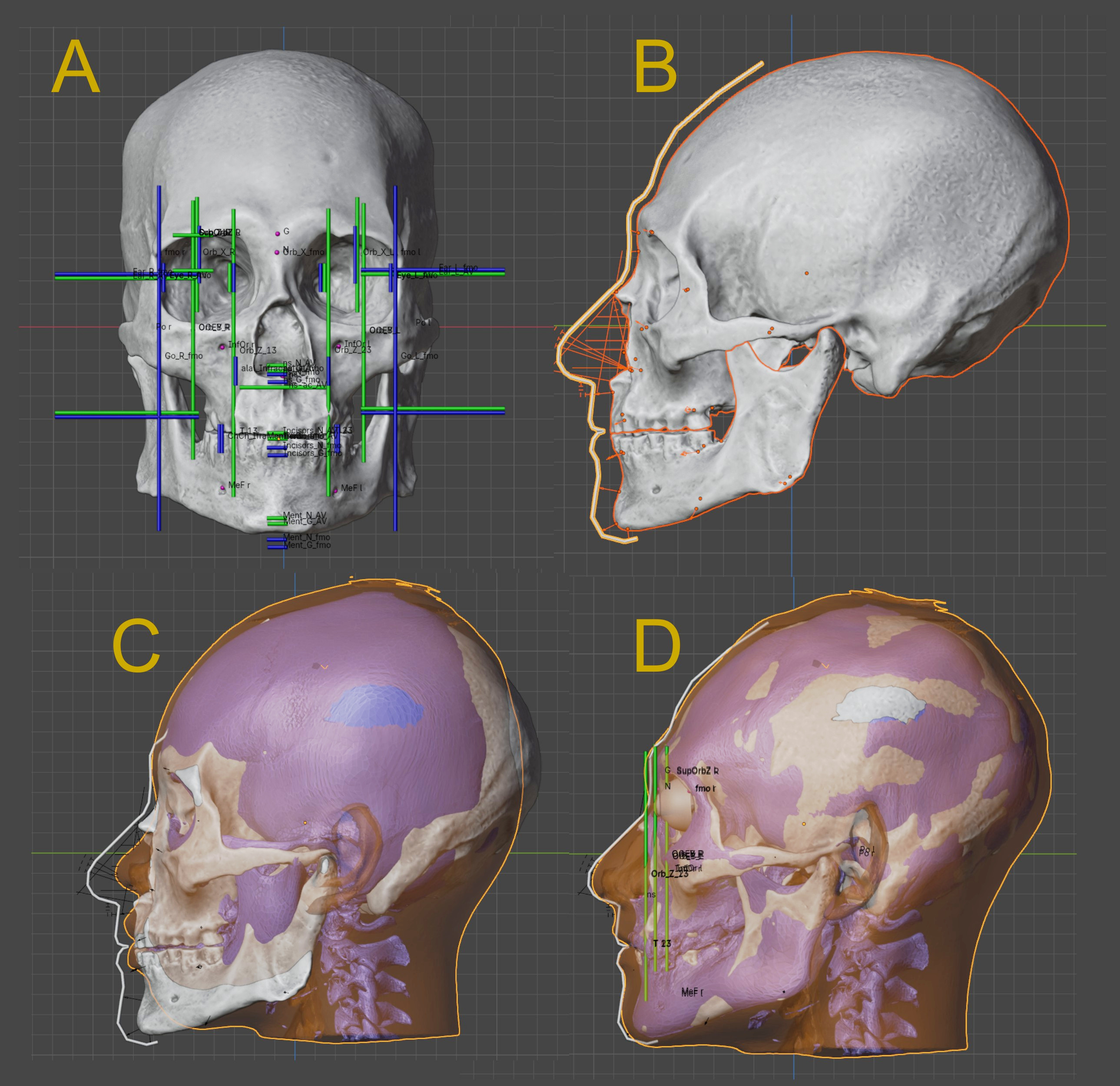
Fig. 1 A) Skull alignet in the Frankfurt plane with projection lines. B) Profile view with soft tissue markers and nose projection. C and D) Facial deformation using a virtual donor.
Initially, two models were downloaded: the skull and the mandible, both provided by the user nicamarvin2005 on Sketchfab. These models are released under the Creative Commons Attribution license. The scale was adjusted using the measurements reported in [Chatters_2000b_a]. The imported model was then aligned in the Frankfurt horizontal plane and subjected to structural projections in OrtogOnBlender, based on data derived from CT scans of living individuals available in [Moraes_and_Suharschi_2022_a]. The teeth of Kennewick Man (hereafter referred to as KM) exhibit considerable wear, resulting in a more closed bite without overbite. This condition causes the facial height to appear slightly reduced along the Z-axis, which is observable in the menton projection: the green lines represent population averages, while the blue lines indicate proportions scaled from the frontomalare orbitale (fmo–fmo) distance. Although the incisor projections align closely with the green (average) lines, suggesting apparent concordance, the correct proportional position corresponds to the lower blue lines. This discrepancy arises because the average fmo–fmo distance is approximately 97 mm, whereas in KM it measures 103 mm—roughly one standard deviation above the mean (https://bit.ly/3NRw2KW). Consequently, the lower limit of the menton falls between the green (average) and blue (proportional) lines. Dental wear of this nature typically produces a modest reduction in facial height along the Z-axis and confers a somewhat prognathic facial appearance (Fig. 1, A).
The line projections indicate the expected positions of key anatomical structures, such as the ocular globes, ears, and mouth, among others. However, to achieve a more accurate facial approximation, additional data are required, including nose projection (derived from the templates in [Moraes_and_Suharschi_2022_a]) and soft-tissue depth markers obtained from a sample of modern Asian individuals [Chan_et_al_2011_a]. By integrating these two datasets, it becomes possible to delineate the facial profile (Fig. 1, B).
It is worth noting that, in the projection model employed here, the Asian reference sample yields a somewhat shorter nasal projection along the Y-axis compared to European samples (see Base AV: https://bit.ly/3NRw2KW). Although these data suffice for a reasonable facial approximation, a further step is undertaken to enhance anatomical detail. This process, known as anatomical deformation [Quatrehomme_et_al_1997_a], involves deforming a virtual donor head—reconstructed from a CT scan of a living individual—until the donor’s skull aligns precisely with that of KM. The resulting deformation is propagated to the soft tissues, producing a final facial approximation that remains consistent with the established projections and profile view (Fig. 1, D and E).
Hint
Didactic materials, in the form of recorded tutorials for OrtogOnBlender, are available as follows: two covering line projections (class 1, class 2), one dedicated to nose projection (here), and one focused on anatomical deformation (here).
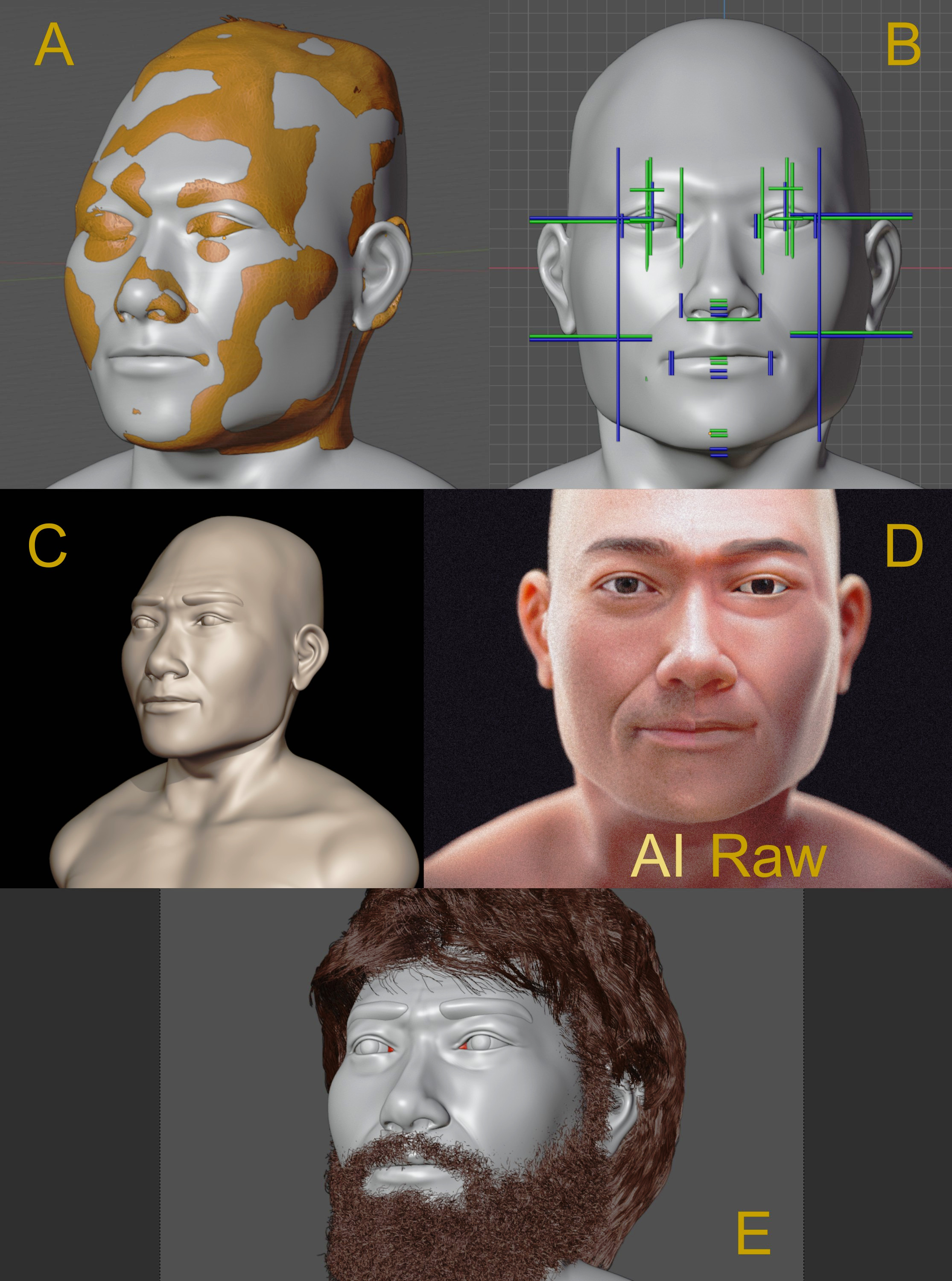
Fig. 2 A) Basic facial structure compatible with the anatomical deformation. B) Frontal view of the basic facial structure with projection lines. C) Basic Bust. D) Facial details enhanced using artificial intelligence.
By integrating data from skull projections, soft-tissue depth markers, and anatomical deformation, a basic facial approximation is generated through interpolation of these sources (Fig. 2, A). The frontal view facilitates verification of compatibility with the projection lines, confirming that the positions of the ocular globes, the limits of the eyes and mouth, and the contours of the nose and ears align appropriately (Fig. 2, A).
An additional noteworthy observation concerns cranial breadth: in most individuals, the bigonial breadth (go–go) is approximately equal to the frontomalare orbitale distance (fmo–fmo). However, in KM, the fmo–fmo measures approximately 103 mm, while the go–go reaches ~120 mm—representing 3.8 standard deviations above the mean. This pronounced bizygomatic and bigonial breadth contributes to the characteristically square facial appearance, which is further accentuated by the reduced facial height along the Z-axis.
The basic facial structure subsequently undergoes refinement through digital sculpting and carving, representing the most objective phase of the approximation process (Fig. 2, C). The final stage involves texturing, coloring, lighting, and fine detailing enhanced by artificial intelligence (Fig. 2, D), employing the Codeformer library [Zhou_et_al_2022_a] running inside OrtogOnBlender. For the artistic implementation, elements of a more conjectural nature, notably fur and hair, were modeled with the intention of producing a distribution characterized by greater naturalness and increased randomness (Fig. 2, E).
Results and Discussion
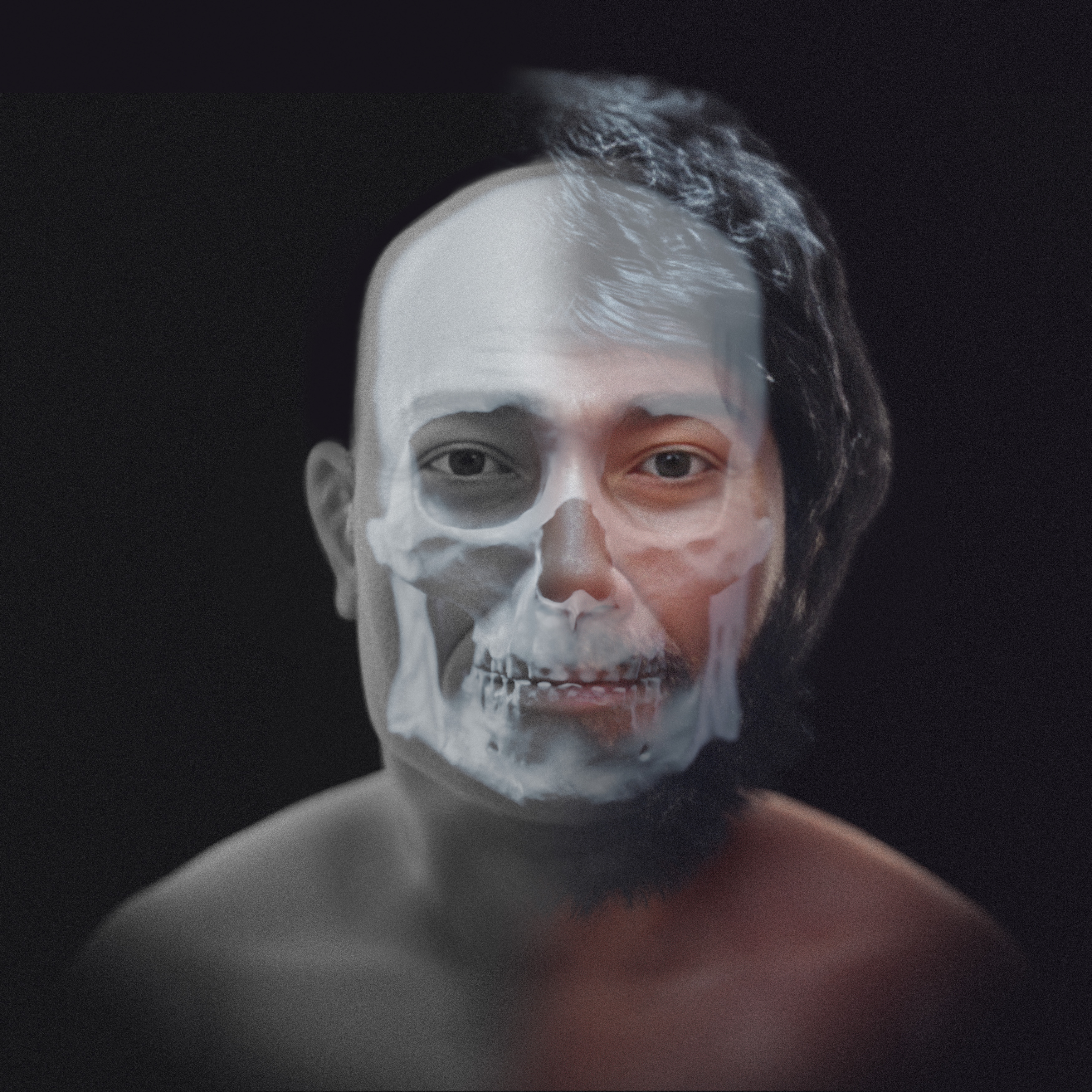
Three distinct groups of images were produced. The first group depicts the most objective and statistically grounded approximation: a basic facial structure in grayscale (Fig. 3). The second group presents rendered faces in grayscale without hair, aligning with conventional forensic imaging standards (Fig. 4, Fig. 5). Finally, the third group features fully colored renderings that incorporate artistic and speculative elements—such as hair and skin tone—informed by the facial approximation published in 2014 (Fig. 6, Fig. 7).
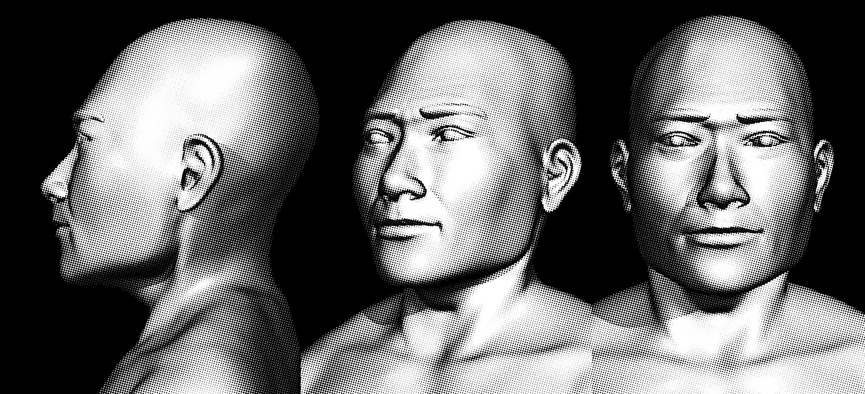
Fig. 3 Basic facial approximation
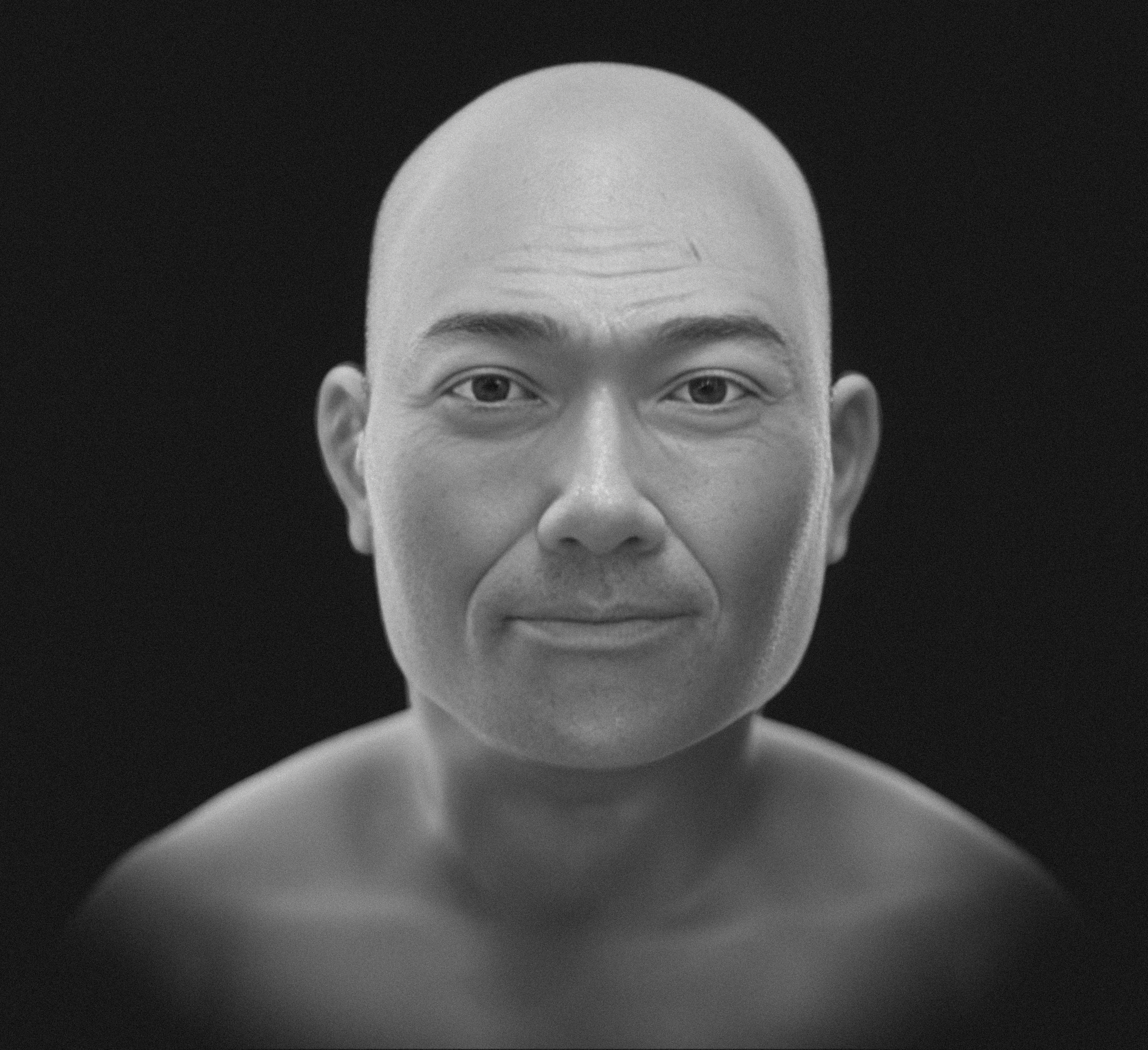
Fig. 4 Simple facial approximation - front
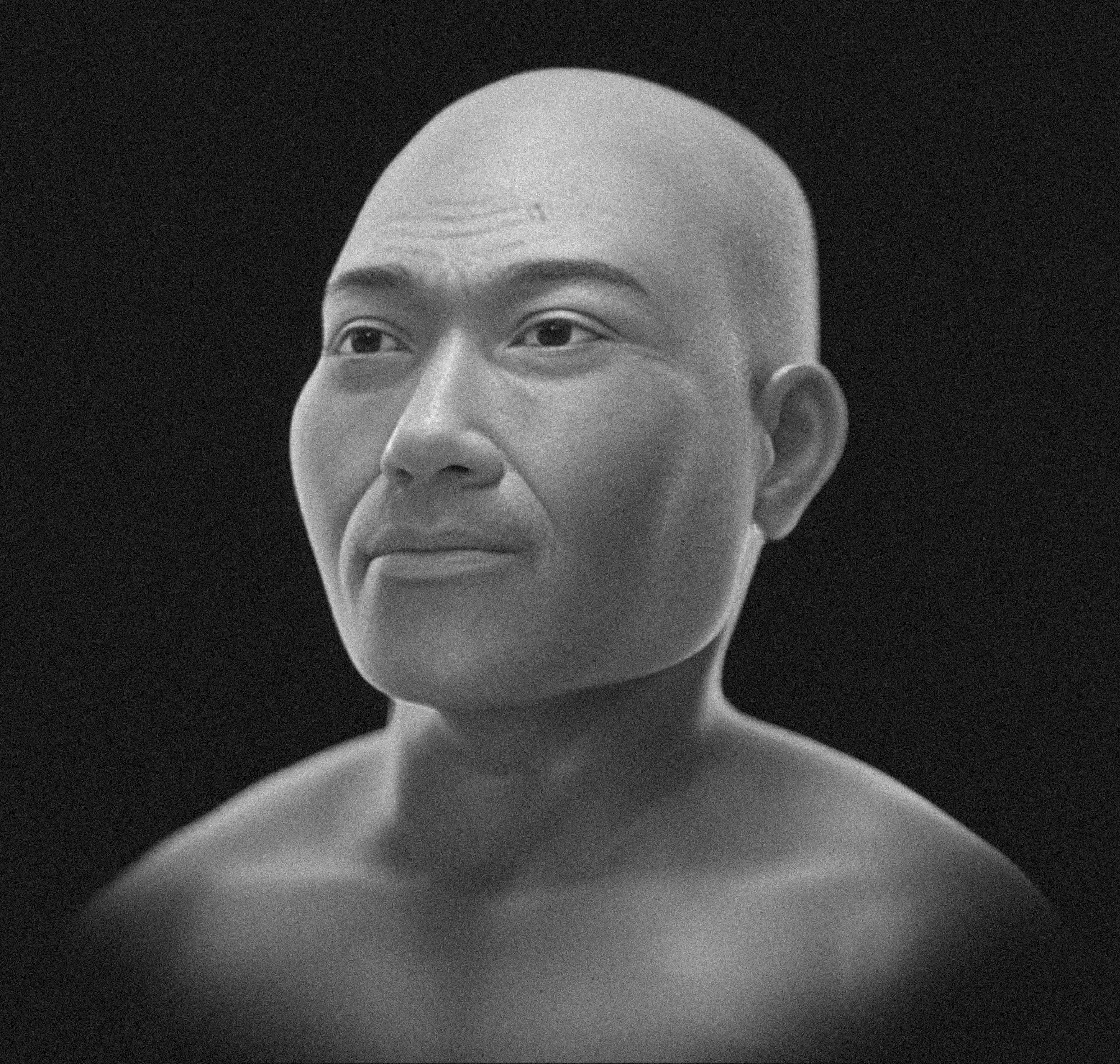
Fig. 5 Simple facial approximation - portrait
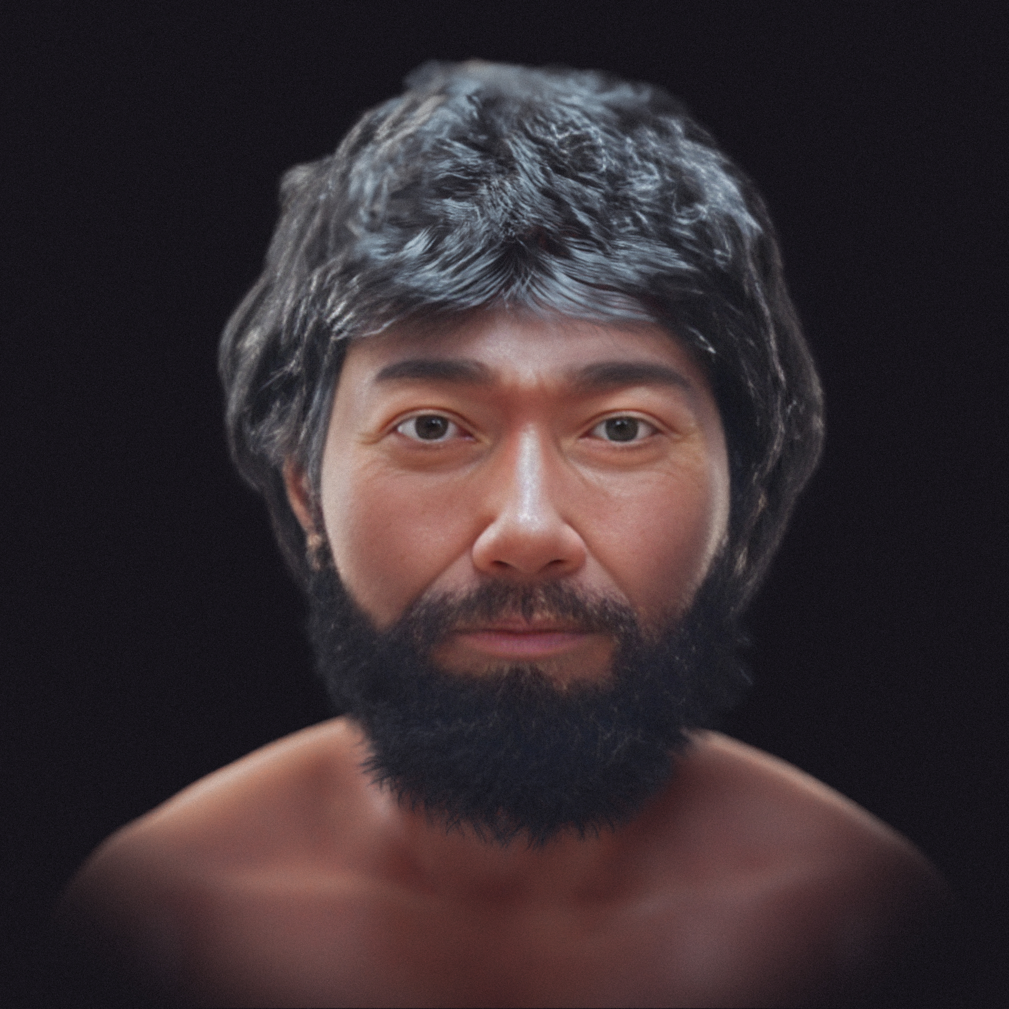
Fig. 6 Artistic facial approximation - front
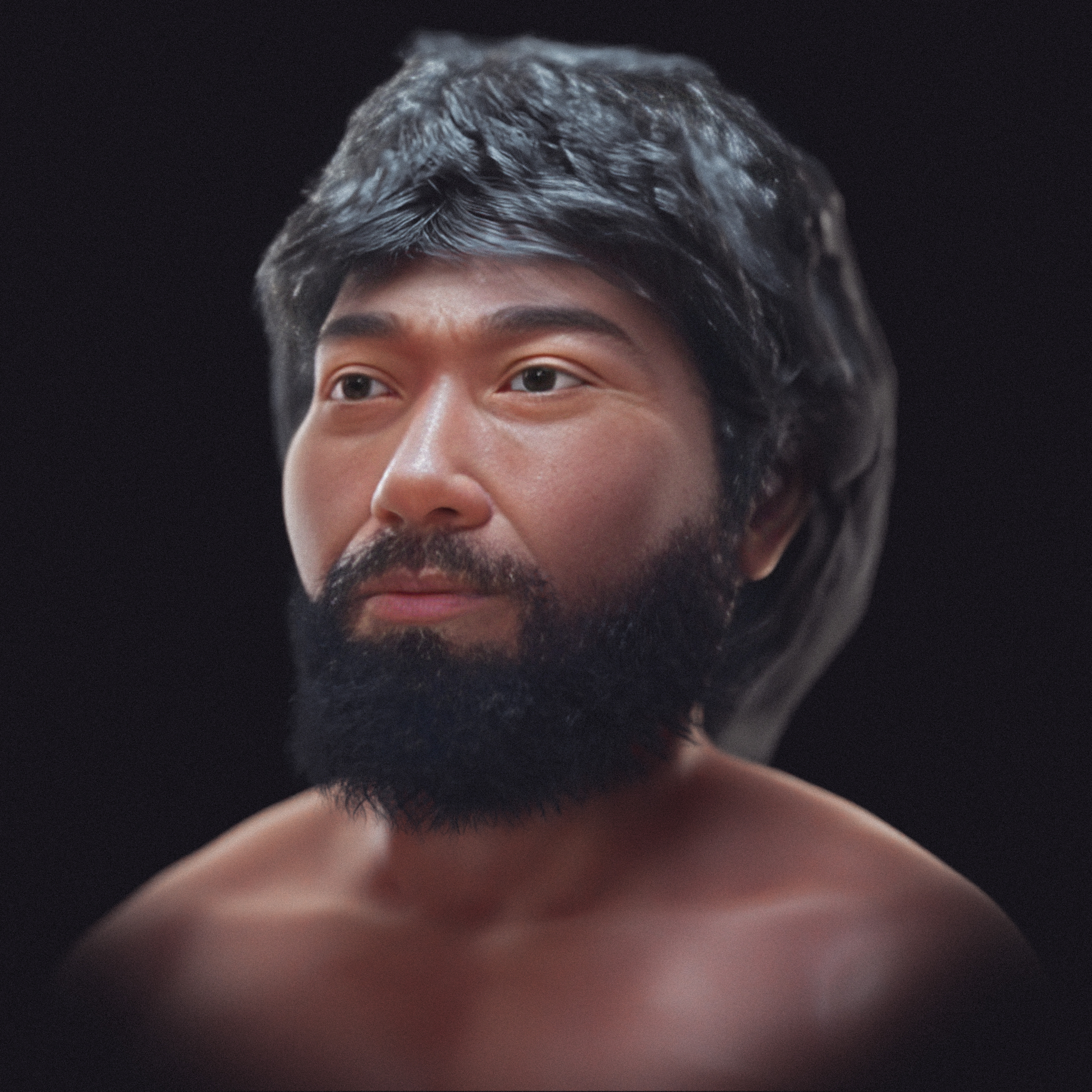
Fig. 7 Artistic facial approximation - portrait
During the anatomical deformation process, the endocast was segmented, yielding an approximate volume of 1450 cm³. When converted to brain volume [Moraes_et_al_2023_a], this corresponds to approximately 1308 cm³—slightly above the modern male average of 1234 cm³ [Ritchie_2018_a]. These measurements should be interpreted with caution. Although they are broadly consistent with the original cranial structure, minor discrepancies may arise between the original publication data and the digitized model, as the mesh reconstruction process can introduce subtle deformations or artifacts.
Conclusion
The facial approximation presented here demonstrates that it is possible to produce technically grounded, reproducible, and transparent forensic reconstructions using open-access data, by integrating anatomical projections, soft tissue markers, and virtual anatomical deformation. By clearly distinguishing between objective results and artistic elements, the study reinforces the scientific and educational value of the methodology without exceeding its inferential limits. This work contributes a didactic model for teaching and research in digital forensic anthropology.
Acknowledgements
To Dr. Dong Quang for providing the tomography of the virtual donor. To nicamarvin2005 (Sketchup user), for share this model and allow this work be done.
Important
This material uses the following Creative Commons license: Attribution 4.0 International (CC BY 4.0).
References
Chan, W. N. J., Listi, G. A., & Manhein, M. H. (2011). In VivoFacial Tissue Depth Study of Chinese‐American Adults in New York City*. Journal of Forensic Sciences, 56(2), 350–358. https://doi.org/10.1111/j.1556-4029.2010.01640.x
Chatters, J. C. (2000). The Recovery and First Analysis of an Early Holocene Human Skeleton from Kennewick, Washington. American Antiquity, 65(2), 291–316. https://doi.org/10.2307/2694060
Chatters, J. (2000). Meet Kennewick Man. NOVA - PBS. https://www.pbs.org/wgbh/nova/article/meet-kennewick-man/
Chatters, J. C. (2017). Making archaeological sense of Kennewick Man. Quaternary International, 444, 83–97. https://doi.org/10.1016/j.quaint.2017.01.045
Mabbutt, L. (2017). Tribes bury remains of ancient ancestor, also called Kennewick Man. The Seattle Times. https://www.seattletimes.com/seattle-news/tribes-bury-remains-of-ancient-ancestor-also-called-kennewick-man/
Moraes, C., & Suharschi, I. (2022). Mensuração de Dados Faciais Ortográficos em Moldavos e Comparação com Outras Populações. figshare. https://doi.org/10.6084/M9.FIGSHARE.20089754
Moraes, C., Habicht, M. E., Galassi, F. M., Varotto, E., & Beaini, T. (2023). Pharaoh Tutankhamun: a novel 3D digital facial approximation. Italian Journal of Anatomy and Embryology, 127(1), 13–22. https://doi.org/10.36253/ijae-14514
Preston, D. (2014). Kennewick Man finally freed to share his secrets. Smithsonian Magazine. https://www.smithsonianmag.com/history/kennewick-man-finally-freed-share-his-secrets-180952462/
Quatrehomme G, Cotin S, Subsol G, Delingette H, Garidel Y, Grévin G, Fidrich M, Bailet P, Ollier A. A fully three-dimensional method for facial reconstruction based on deformable models. J Forensic Sci. 1997 Jul;42(4):649-52. PMID: 9243826.
Rasmussen, M., Sikora, M., Albrechtsen, A., Korneliussen, T. S., Moreno-Mayar, J. V., Poznik, G. D., Zollikofer, C. P. E., Ponce de León, M. S., Allentoft, M. E., Moltke, I., Jónsson, H., Valdiosera, C., Malhi, R. S., Orlando, L., Bustamante, C. D., Stafford, T. W., Jr, Meltzer, D. J., Nielsen, R., & Willerslev, E. (2015). The ancestry and affiliations of Kennewick Man. Nature, 523(7561), 455–458. https://doi.org/10.1038/nature14625
Ritchie, S. J., Cox, S. R., Shen, X., Lombardo, M. V., Reus, L. M., Alloza, C., Harris, M. A., Alderson, H. L., Hunter, S., Neilson, E., Liewald, D. C. M., Auyeung, B., Whalley, H. C., Lawrie, S. M., Gale, C. R., Bastin, M. E., McIntosh, A. M., & Deary, I. J. (2018). Sex Differences in the Adult Human Brain: Evidence from 5216 UK Biobank Participants. In Cerebral Cortex (Vol. 28, Issue 8, pp. 2959–2975). Oxford University Press (OUP). https://doi.org/10.1093/cercor/bhy109
Zhou, S., Chan, K. C. K., Li, C., & Loy, C. C. (2022). Towards Robust Blind Face Restoration with Codebook Lookup Transformer (Version 2). arXiv. https://doi.org/10.48550/ARXIV.2206.11253