OrtogOnBlender: Radical Transparency, Open Science, and Conceptual Alignment with Regulatory Frameworks
Attention
This document does not constitute, nor does it claim to constitute, a de jure regulatory certification process; it is a voluntary exercise of transparency and conceptual alignment with internationally recognized formal protocols.
Abstract
This paper presents OrtogOnBlender (OOB), an open-source, free-software (FLOSS) add-on designed for virtual surgical planning. Conceptually aligned with international regulatory guidelines, OOB functions de facto as a Software as a Medical Device (SaMD) under Class II risk categorization. Although officially launched as a unified ecosystem in 2017, its technological core represents over a decade of clinical and scientific incubation. This developmental background spans early research in forensic facial approximation, low-cost submillimeter photogrammetry, and pioneering 3D-printed veterinary prostheses, establishing a highly resilient mathematical foundation. To address safety and reproducibility requirements, OOB enforces a strict configuration control by freezing its environments across stable versions (OOB 291 and OOB XP), mitigating risks associated with external software dependencies (SOUP). To actively counter the “black-box” paradigm of proprietary medical suites, the project operates under a philosophy of radical transparency, offering open access to its complete Python source code and algorithm databases. Furthermore, the system is designed around a “human-in-the-loop” model: OOB does not perform autonomous medical decision-making; rather, it is operated under the strict supervision, verification, and final approval of the surgeon. A comprehensive 10-step fiduciarily crossed validation protocol—integrating CT scan voxel data, intraoral scans, and ArUco-calibrated photogrammetry—empowers the clinical operator to actively audit and confirm structural accuracy before physical manufacturing. Supported by a continuous publishing model through the OrtogOnLineMag and numerous peer-reviewed studies, OOB demonstrates how open science can foster reliable, auditable, and globally cooperative medical planning, reaching active users across 38 countries.
Important
Due to its strictly dynamic and documentary nature, this material is subject to periodic reviews, methodological updates, and continuous technical refinements throughout its life cycle. The present reference version is consolidated and indexed under the configuration control temporal registry 20260714.
Introduction and History
Background
The development of OrtogOnBlender (OOB) consolidates an accumulation of technical and scientific expertise developed by the authors over more than a decade. The project’s history reflects the gradual transition from tools initially conceived in an open-source environment with wide media coverage to a methodologically mature ecosystem integrated into the academic field. In 2011, one of the authors initiated investigations in the field of forensic facial reconstruction using Blender—a free and open-source 3D modeling and animation software—in convergence with InVesalius (https://invesalius.github.io/), which focuses on the three-dimensional reconstruction of DICOM tomographic files. One of the first studies derived from this workflow, published in 2012, indicated the theoretical feasibility of combining these tools for the design of customized craniofacial prostheses. In 2013, the development of a photogrammetry protocol aimed at the three-dimensional digitization of skeletal elements [Dias_et_al_2013_a] achieved metric equivalence to structured light systems, receiving first place as a scientific poster presentation (Fig. 1) at an official Forensic Dentistry event of the University of São Paulo (USP).
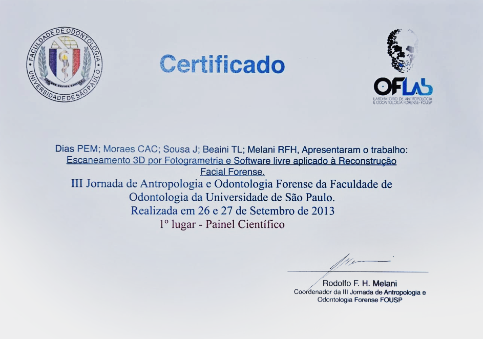
Fig. 1 Scientific poster award certificate - 1st Place (2013).
In 2014, the results obtained from forensic facial approximation research reached national and international media prominence. This milestone drew the interest of the other two co-authors of the add-on, who identified in the methodology the potential to optimize virtual surgical planning. Although the specialists had no prior contact with one another, they parallelly initiated training focused on adapting these computational resources to surgical and biomedical routines. This technical-clinical cooperation highlighted the necessity of implementing more robust and predictable engineering solutions, since Blender’s native engine at the time lacked optimized geometric tools for surgical demands. Consequently, the integration of third-party algorithms and libraries was pursued to guarantee the millimeter-level precision of the processes.
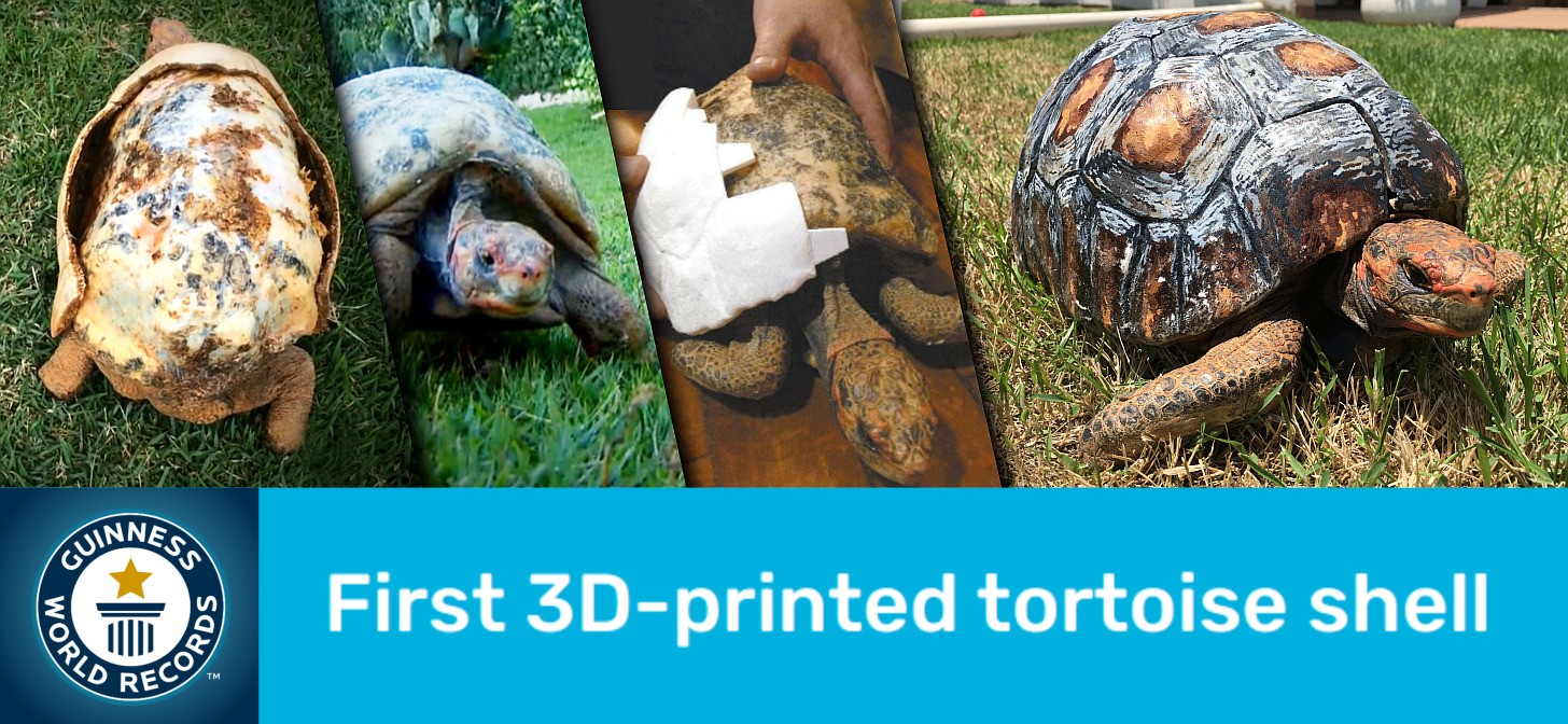
Fig. 2 Guinness World Record recognition for the first 3D-printed tortoise shell in the world.
The integration of external libraries specialized in complex boolean calculations, such as Cork (https://github.com/gilbo/cork), in combination with the photogrammetry pipeline initially based on PPT-GUI [Moulon_and_Bezzi_2011_a], expanded the protocol’s field of application to reconstructive veterinary medicine. This synergy enabled the design and additive manufacturing of the world’s first 3D-printed wildlife prostheses, including specimens such as a toucan and a tortoise [Rabello_et_al_2016_a]—an achievement internationally registered by the Guinness World Records (Fig. 2).
Concurrently with technical advancements, the surgical protocol was validated in multiple complex clinical cases, though it still relied on the fragmented execution of separate software programs. In 2016, presentations and lectures at technology events publicly documented the accuracy and practical application of these customized surgical cutting guides, a methodology that also became established in complex veterinary interventions.
Clinical demand expanded toward the manufacture of high-complexity facial prostheses. This field required maximum fidelity from photogrammetry to capture soft tissue texture and relief, alongside high computational performance from boolean algorithms to preserve fine anatomical surface details in the digital environment. In order to disseminate the accumulated knowledge, the authors structured technical training courses. However, the need to manipulate a fragmented suite of independent software programs imposed a steep learning curve and high operational fatigue on students due to the sheer volume of different commands required to complete a single virtual plan.
The Development of OrtogOnBlender
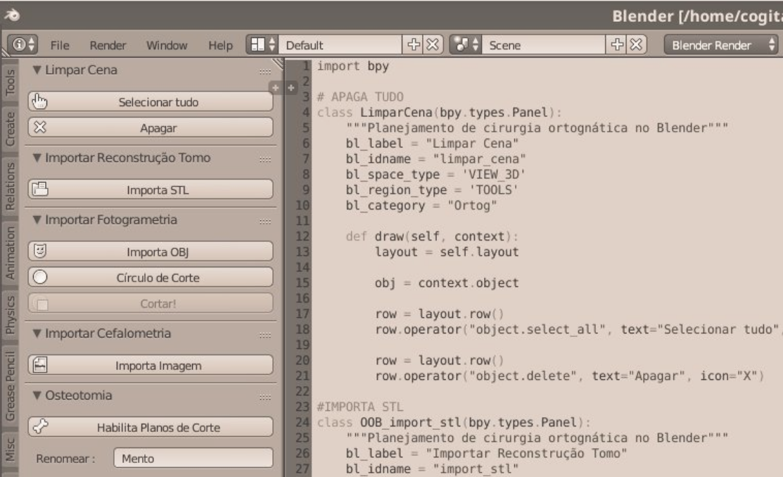
Fig. 3 First screenshot of the OOB interface in September 2017.
Based on the mapping of the main operational difficulties reported by students, an initial set of scripts and macro tools was developed in 2017 to automate bureaucratic and redundant tasks. This initial core grouped routines such as global scene cleanup, standardized import of generic files in .OBJ (derived from photogrammetry) and .STL (generated by tomographic reconstructions) formats, as well as the parameterized insertion of osteotomy cutting planes (Fig. 3). Although the component was characterized as a low abstract complexity implementation, the reception by new students and veteran users was highly favorable due to the immediate optimization of the workflow.
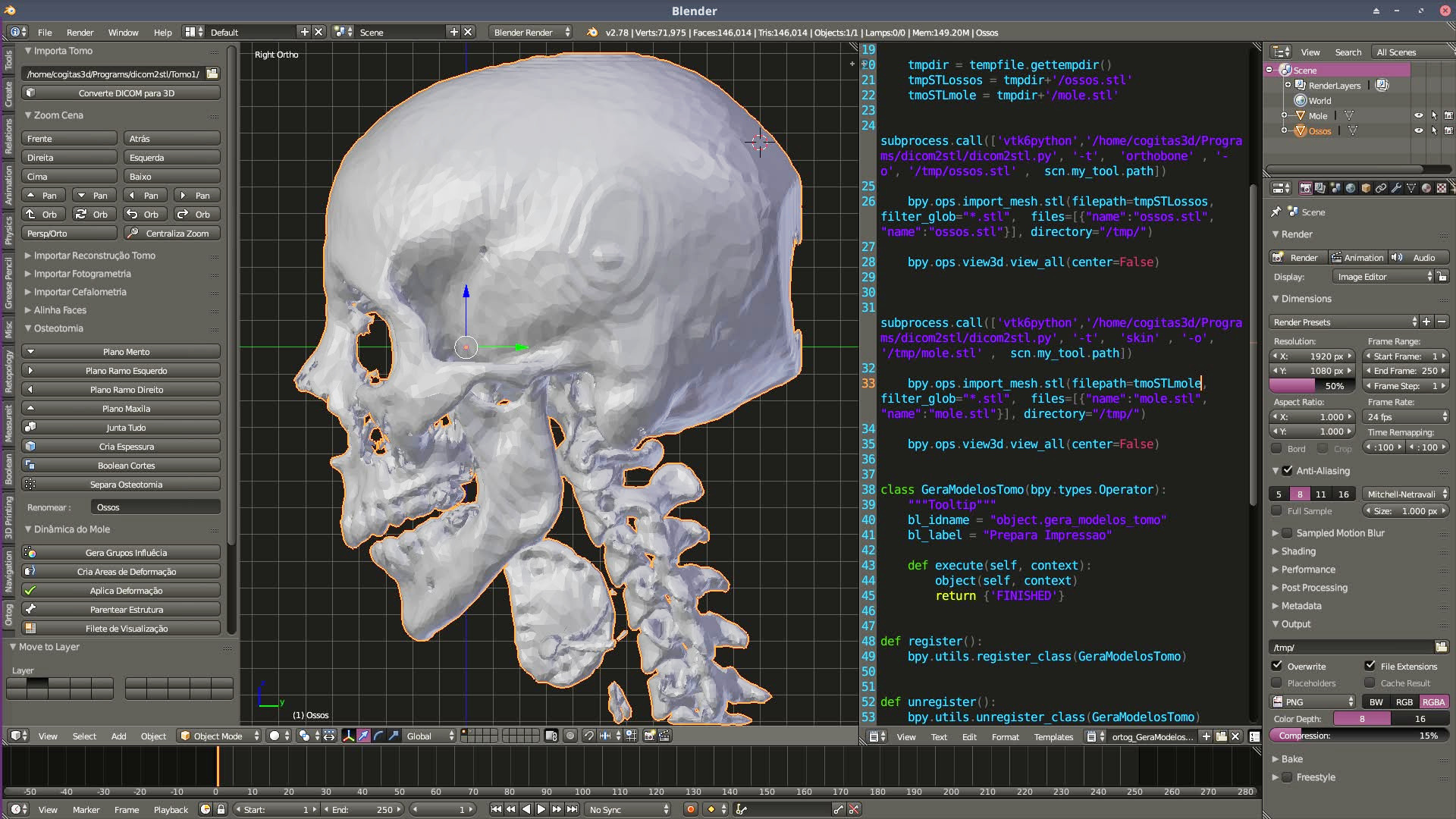
Fig. 4 First screenshot of a computed tomography (CT-Scan) reconstruction test on November 30, 2017.
The consolidation of this interface and the subsequent increase in productivity motivated, approximately two months after the initial launch, the development and testing of routines for the direct import and native conversion of computed tomography (CT-Scan) data (Fig. 4). However, although preliminary experiments were successful in controlled environments, a significant portion of the tools failed when subjected to stress tests with real-world examinations. These inconsistencies stemmed from the wide heterogeneity of tomograph manufacturers, variations in voxel matrix dimensions and spacing (voxel data), fluctuating Hounsfield scales, and spatial orientation distortions. In this technical scenario, the first exogenous tool to demonstrate mathematical stability and reproducibility in mesh rendering tests was the open-source library DicomToMesh (https://github.com/eidelen/DicomToMesh).
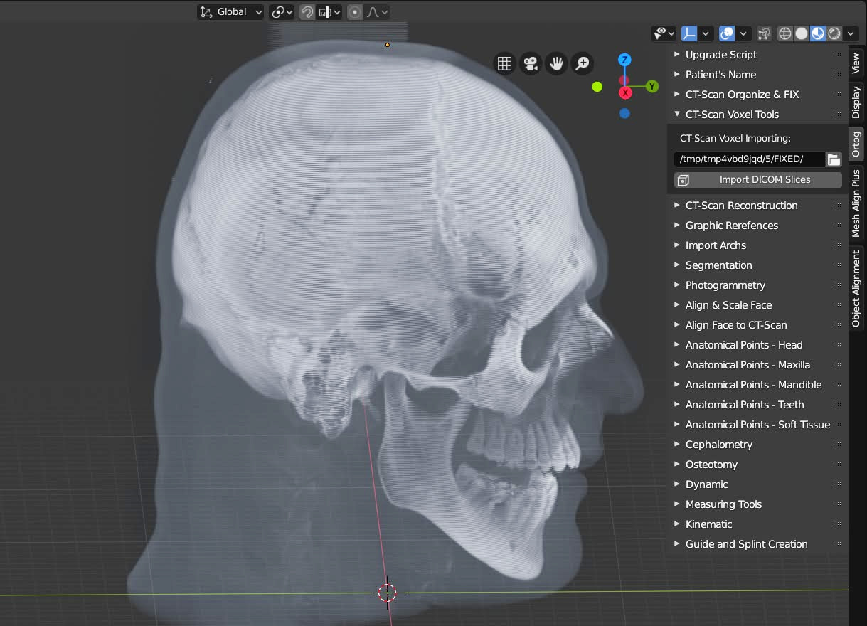
Fig. 5 First tool for importing and displaying voxel data.
Despite the fact that the isomorphic three-dimensional meshes extracted from tomographies provided the necessary geometric data for virtual surgical planning, specialists frequently requested comparative visual inspection of the original tomographic slices, given that raw volume data (voxel data) preserved a substantially richer structural detail of tissue density. To bridge this gap, a volumetric data import system was implemented which, in its early phase, allowed the spatial projection of the mapped tomographic block strictly in grayscale (Fig. 5). At this evolutionary stage of the add-on, the execution of essential virtual planning tasks for orthognathic surgery still depended on text-mode CLI (Command Line Interface) calls to subordinate external routines, such as the Cork library for complex bone boolean calculations in osteotomies, the DicomToSTL pipeline for the three-dimensional reconstruction of DICOM files, and the ImageMagick software (https://imagemagick.org/) for converting DICOM slices into rasterized image sequences, serving as the basis for voxel data reconstruction in Blender.
The progressive and automated integration of these tools into the add-on’s source code eliminated the need for the operator to manually resort to multiple external programs, consolidating the ecosystem as a self-contained solution for virtual planning in craniomaxillofacial surgery.
Community
The creation of the official OOB user group on the WhatsApp platform occurred on September 1, 2017, with the initial objective of concentrating students and operators of the ecosystem. As will be discussed in detail in subsequent sections, the early grouping of these professionals made it possible to establish a direct channel for immediate communication. This open governance strategy guaranteed that the add-on’s development cycle was systematically guided and prioritized by the clinical demands and real practical needs expressed by the user community.
Submodules
The demographic profile of the OOB community is characterized mainly by surgeons and technicians specialized in virtual surgical planning workflows. Due to the multidisciplinary nature of this technical body, operational demands quickly transcended the exclusive scope of classic orthognathic surgery. Faced with this necessity, the add-on underwent a process of modular and architectural expansion, culminating in the conception of dedicated submodules integrated into the main core:
RhinOnBlender: Developed specifically for the three-dimensional simulation and virtual planning of rhinoplasties;
ForensicOnBlender: Optimized for forensic facial reconstruction and approximation routines from archaeological or forensic skeletal remains;
+IDOnBlender: Directed toward the digital design and manufacturing of customized facial prostheses for maxillofacial rehabilitation;
OthersOnBlender: Configured as a laboratory testing environment for new tools and scripts in the experimental phase which, if consistently validated and absorbed by the community, are permanently ported to the corresponding submodules or consolidated into new standalone extensions.
Documentation
In 2019, the official OrtogOnBlender documentation was centralized in a permanent digital repository (https://www.ciceromoraes.com.br/doc/pt_br/OrtogOnBlender/index.html), where it remains available. The scope of this documentary collection ranges from the fundamental concepts of applied computer graphics to the installation routines of the stable OOB 291 version in Windows, macOS, and GNU/Linux enterprise environments. Additionally, the material covers clinical protocols for medical radiological image acquisition, facial photogrammetry guidelines, and laboratory test reports focused on measuring the three-dimensional submillimeter precision of the ecosystem.
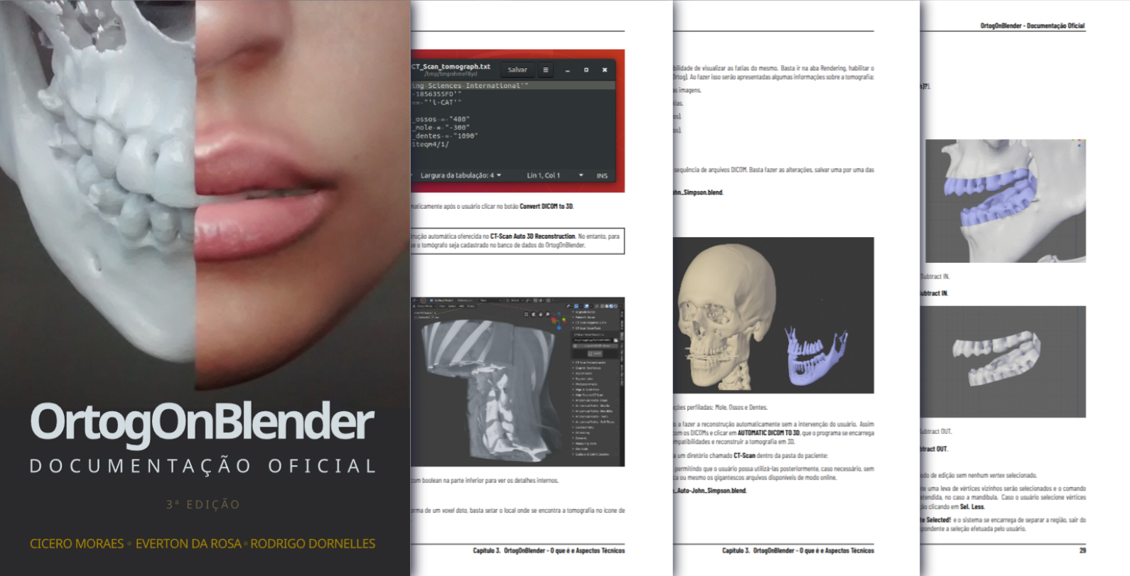
Fig. 6 PDF version of the third edition of the official OOB documentation.
The progressive volume of chapters dedicated to algorithmic stress tests and structural accuracy evaluations substantially expanded the scope of the original technical documentation, temporarily distancing it from the strict format of a standard installation manual. Faced with this accelerated growth and the need to document the continuous evolution of the source code, the technical-scientific journal OrtogOnLineMag was founded in 2020. The journal operates coordinately as an extension of the add-on’s official documentation, functioning as a dynamic repository to record the development of new scripts, biomedical trials, and practical applications of the ecosystem.
Note
The educational background of the primary developer of OOB in the field of computing dates back to the second half of the 1990s. During this period, the teaching of operating systems prioritized text-mode interaction via MS-DOS, preceding the transition to graphical user interfaces based on the Windows 3.11 and Windows 95 platforms. In 2005, the developer migrated to the Linux environment, whose logical architecture reinforced the initial command-line learning over subordinate graphical interfaces. This structural foundation is directly reflected in the software architecture of OOB, where multiple tools operate in the background via CLI, with the resulting geometric outputs being seamlessly imported into the graphical viewport. This methodological approach enables the rapid integration of cutting-edge libraries into the ecosystem, as the development of complex scientific tools frequently demands native compilation and text-mode manipulation under multiple operating systems. As a reflection of this structural optimization, the add-on exhibits superior computational performance in Linux environments. Consequently, the official documentation incorporated dedicated tutorials for deploying the portable Linux 3DCS system (based on Ubuntu) on USB flash drives (https://www.ciceromoraes.com.br/doc/pt_br/OrtogOnBlender/Instalacao_Linux3DCS.html) or external solid-state drives (https://www.ciceromoraes.com.br/doc/pt_br/OrtogOnBlender/Instalacao_Linux3DCS_SSD.html), allowing the immediate and self-contained execution of the 3D suite on USB 3.0 buses of any computer, bypassing the need for installation on the local hard drive. There are plans to update the project to the LTS Ubuntu 24.04 distribution, whose extended support cycle of up to 10 years aims to ensure the sustainability of new segmentation technologies based on artificial intelligence.
Formal Publications
As OOB consolidated itself as a de facto solution for virtual surgical planning, the technical rigor associated with the development and maintenance of its tools expanded proportionally. The widespread adoption of the system by surgeons, educators, and university students drove a continuous process of scientific formalization, resulting in the publication of articles in conference proceedings and peer-reviewed international journals.
The inaugural records in the scientific literature citing OOB occurred in 2018, less than a year after the beginning of its development. These initial publications consisted of expanded abstracts presented at oral and maxillofacial surgery and traumatology events, addressing virtual surgical simulation [Souza_et_al_2018_a] and digitization by photogrammetry [Lima_et_al_2018_a], in addition to a peer-reviewed article dedicated to analyzing the operational difficulties experienced by novice users when facing the 3D modeling interface [Shibasaki_et_al_2018_a].
In 2019, the first international abstract was published in an indexed supplement, attesting that the ecosystem provided the necessary geometric support for the stable execution of complex surgical planning [Pierri_et_al_2019_a].
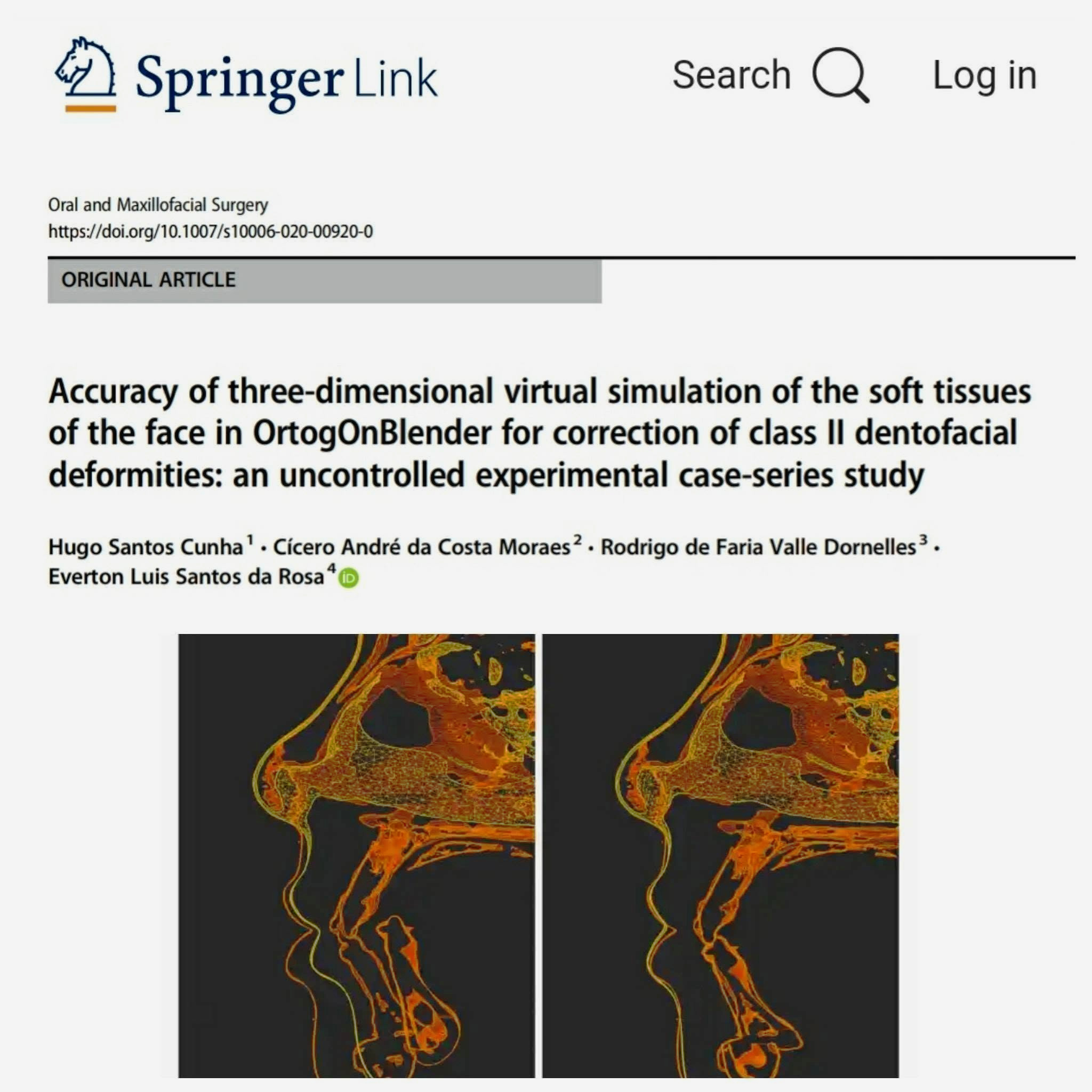
Fig. 7 Original article published in the journal Maxillofacial Surgery (Springer Nature).
The first peer-reviewed article in a journal with an impact factor was published in 2020. The study compared a series of digitally planned cases with their respective post-operative results, demonstrating a statistically significant structural compatibility situated well within the metric tolerance margins established by clinical literature [Cunha_et_al_2020_a]. Although the aforementioned article counted on the co-authorship of the tool’s developers—an aspect open to discussions regarding potential conflicts of interest, despite the technology being strictly free and open-source—an independent study conducted by external researchers and published in 2022 not only corroborated the original accuracy rates, but also demonstrated statistical equivalence between OOB and a high-cost proprietary software widely consolidated in the medical market [Lobo_et_al_2022_a].
Important
By virtue of its open architecture and free license, OOB is frequently employed in a complementary and integrated manner with proprietary software ecosystems, including those dedicated to virtual surgical planning. Multiple specialists trained in this domain carry out the methodological convergence between the commercial tools of their expertise and the wide range of additional features made available by OOB, such as the fabrication of customized surgical guides, photogrammetry pipelines, and anatomical segmentation routines based on artificial intelligence. Thus, the scope of OOB is not characterized by hostile competition against other market solutions, but rather by functional synergy, valuing the specialist’s prior clinical knowledge and providing technological support to optimize the execution of their professional practice.
Within the specialized submodules of the ecosystem, the pioneer in scientific publications was RhinOnBlender, presented as a conference paper in 2018 during an international conference on health-oriented computer graphics [Dornelles_et_al_2018_a]. Subsequently, the first peer-reviewed case report of this module was accepted in the Aesthetic Surgery Journal, classified at the time as one of the main global references in plastic surgery (#2) and aesthetics (#1) [Sobral_et_al_2021_a].
Also in 2021, a case report applying the core of the add-on in a distinct area was published, documenting the effectiveness of the tool in the planning and guided reduction of severe atrophic mandibular fractures [Facanha_25_al_2021_a].
The +IDOnBlender submodule obtained its first peer-reviewed article in 2021, focusing on the efficiency of the tool applied to facial prosthetic rehabilitation [Falih_et_al_2021_a]. In 2022, the digital design protocol of this module demonstrated technical maturity by validating new direct additive manufacturing workflows for facial prostheses in photopolymerizable resin, bypassing the analog laboratory stages of negative impression molding [Salazar_et_al_2022_a]. Finally, the ForensicOnBlender submodule had its inaugural peer-reviewed article accepted in 2022, consolidating the metric precision of the add-on’s algorithms applied to facial approximation methodologies from archaeological remains [Abdullah_et_al_2022_a].
Currently, the scientific literature linked to the OOB ecosystem is widely indexed in highly prestigious international databases, such as PubMed, Google Scholar, and the like, ratifying the project’s commitment to academic validation and the methodological transparency of its results.
Geographical Distribution
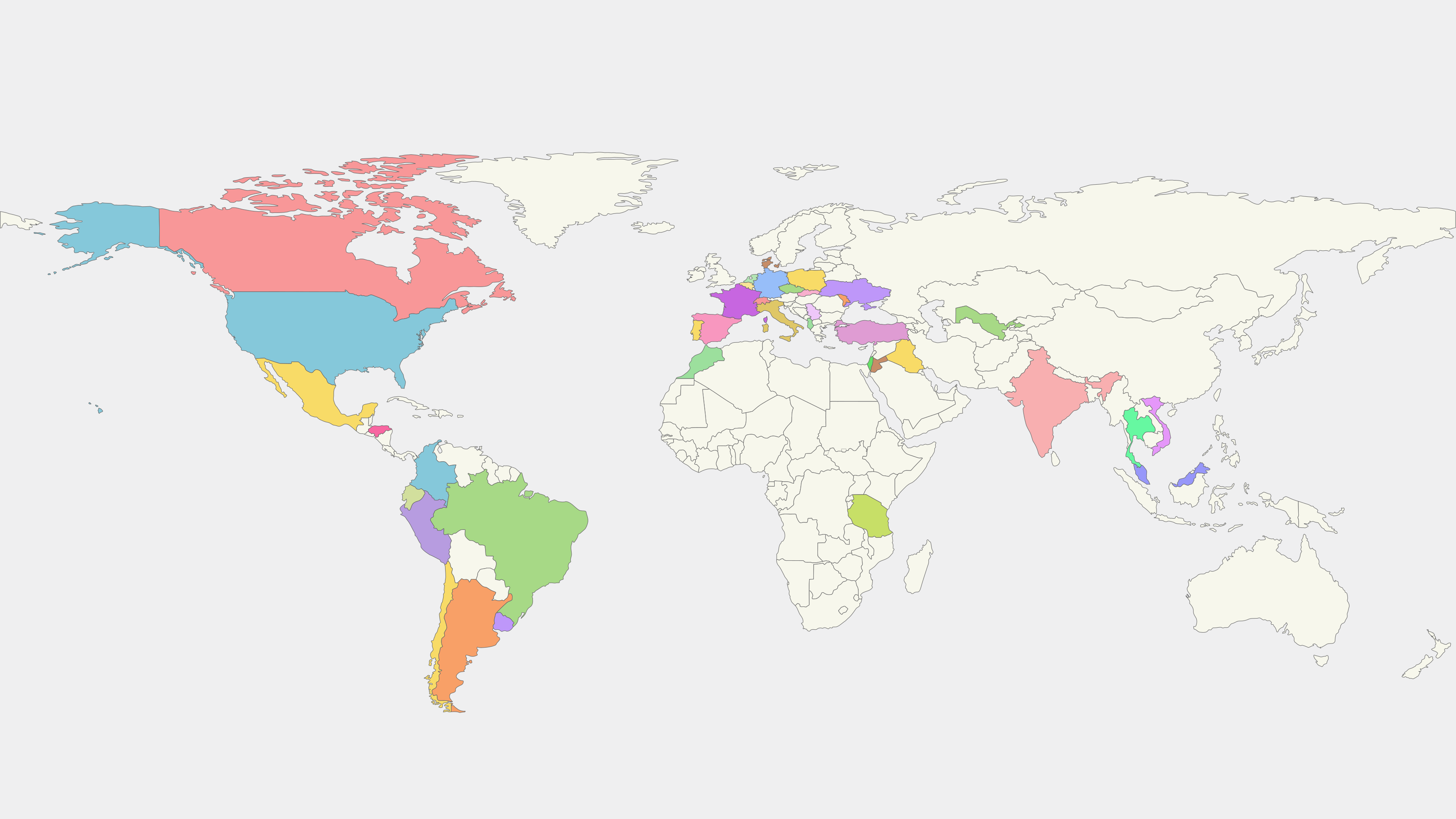
Fig. 8 Countries with registered users.
The survey of countries with OOB users is based on the nationality and international country calling codes (DDI) of the participants in the official WhatsApp group for the script; thus, the actual number may be higher[cite: 1]. Currently, there are active users in 38 countries (Fig. 8), namely: Albania, Argentina, Belgium, Brazil, Canada, Chile, Colombia, Czech Republic, Denmark, Ecuador, France, Germany, Honduras, India, Iraq, Israel, Italy, Jordan, Malaysia, Mexico, Moldova, Morocco, Netherlands, Peru, Poland, Portugal, Serbia, Slovakia, Spain, Switzerland, Tanzania, Thailand, Türkiye, Ukraine, United States, Uruguay, Uzbekistan, and Vietnam.
Users voluntarily provided the names of the hospitals, clinics, and institutions where they work or participate, including: ADN Hospital in Andijan City – Andijan City – Uzbekistan, Alalwya Specialized Dental Centre – Iraq, Alliance Hospital – Brazil, Armed Forces Hospital – Brasília – Brazil, Avisenna Specialist Hospital – Malaysia, Base hospital of the Federal District – Brasília – Brazil, Basic Hospital of San Marcos Ocotepeque – San Marcos Ocotepeque – Honduras, Beneficência Portuguesa Hospital – São Paulo – Brazil, Cajuru University Hospital – Curitiba – Brazil, Cardiopulmonary Hospital – Brazil, CEMA São Paulo Hospital – São Paulo – Brazil, Dental Polyclinic of the Military Fire Department of the Federal District – Brasília – Brazil, Erasto Gaertner Hospital - Cancer Center – Curitiba – Brazil, Hospital Bom Jesus Vilhena-RO – Vilhena – Brazil, Hospital Check Up – Manaus – Brazil, Hospital Coopera UNIMED Vilhena-RO – Vilhena – Brazil, Hospital das Clínicas (HCFMUSP) – São Paulo – Brazil, Hospital das Nações – Curitiba – Brazil, Hospital Erasto Gaertner - Câncer Center – Curitiba – Brazil, Hospital Regional de Vilhena-RO – Vilhena – Brazil, Hospital Santiago Oriente – Dr. Luis Tisné Brousse – Santiago – Chile, Hospital Universitário da Universidade Estadual de Londrina – Londrina – Brazil, IMSP Institutul de Medicină Urgentă – Moldova, Itaigara Memorial Hospital – Brazil, Jorge Valente Hospital – Brazil, Manaus Adventist Hospital – Manaus – Brazil, Marcelino Champagnat Hospital – Curitiba – Brazil, Mato Grosso Cancer Hospital – Cuiabá – Brazil, Mechnikov Hospital Dnipro – Dnipro – Ukraine, National Hospital of Odonto-Stomatology in Hanoi – Hanoi – Vietnam, National Institute of Neoplastic Diseases – Peru, National Niño Institute of San Borja – San Borja – Peru, Paraná Institute of Otorhinolaryngology – Curitiba – Brazil, Policlinico di Bari – Bari – Italy, Portuguese Hospital of Bahia – Brazil, Santa Casa de Alfenas – Alfenas – Brazil, Santa Casa de Misericórdia de Araçatuba and Unesp – Araçatuba – Brazil, Santa Isabel Hospital – Brazil, Santa Julia Hospital – Manaus – Brazil, Santo Amaro Hospital – Brazil, Santo Antônio Hospital - Sister Dulce Social Works – Brazil, Scientific University of the South – Peru, State Medical University of Medicine and Pharmacy “Nicolae Testemitanu” – Chisinău – Moldova, University Clinical Hospital in Opole – Opole – Poland, Valencia University Clinical Hospital – Valencia – Spain, Wolfson Medical Center – Holon – Israel, České Budějovice Hospital – České Budějovice – Czech Republic.
Technical Aspects
Version History
The initial iterations of OOB were structured on the APIs of Blender versions 2.78 and 2.80. The first frozen version established with permanent release status was OOB 291, coupled with the immutable Blender 2.91 line. Subsequently, in 2024, the cutting-edge development environment was stabilized and frozen on Blender version 4.2, giving rise to OOB XP. The strategic decision to freeze the operating environment aims to guarantee greater reliability and predictability for the biomedical tool, optimizing exception monitoring and bug tracking. Additionally, this engineering approach enables the maintenance of fixed and resilient technical documentation, mitigating the need for constant refactoring with each release cycle of the base platform. In line with the Blender core, the other exogenous components of the graphics suite also undergo strict configuration control and freezing, ranging from subordinate image processing software, such as Slicer, to critical scientific libraries, such as VTK, Torch, CUDA, and related frameworks.
Formalization
The developed scripts and solutions were registered with the INPI - National Institute of Industrial Property of Brazil, under the following process IDs:
The official database containing these public records can be accessed directly at [INPI_2026_a].
Academic Incubator
As discussed preliminarily, the OOB ecosystem is supported by a robust documentation infrastructure, split between fixed normative manuals—focused on installation, configuration, and stable operation guidance of the releases—and the dynamic collection linked to the technical-scientific journal OrtogOnLineMag (OOLM). However, OOLM transcends the role of a mere documentary extension of the project, functioning as an incubator for biomedical techniques and academic publications. Since its founding in 2020, the journal has recorded 14 uninterruptedly published issues, totaling 81 original scientific articles. A significant portion of this editorial ecosystem evolved into consolidated versions in peer-reviewed, indexed international journals, or became trustworthy methodological references in specialized medical literature.
In strict symmetry with the open-source philosophy of OrtogOnBlender, OOLM adopts Open Science guidelines in its production workflow. The editorial pipeline utilizes the Sphinx (https://www.sphinx-doc.org/) documentation framework for plain-text formatting, whose source files are compiled natively into PDF (via LaTeX typesetting) and interactive HTML distributions. Finally, the knowledge generated and distributed is protected and licensed under the terms of the Creative Commons Attribution 4.0 International license (CC BY 4.0).
Structural Precision and Confidence
The two aforementioned items complement each other, since—as a tool used de facto for virtual surgical planning under the strict responsibility of surgeons—it becomes imperative to offer a database centered on informational reliability. In this regard, the trials and articles shared in OOLM, subsequently validated by peer-reviewed journals, empirically ground this technical predictability.
For the practical feasibility of using any software, its deployment must occur in an accessible and standardized manner. This requirement is extensively documented in the stable release version (OOB 291), which provides detailed step-by-step guidelines for the Windows (https://www.ciceromoraes.com.br/doc/pt_br/OrtogOnBlender/Instalacao_Windows.html), macOS (https://www.ciceromoraes.com.br/doc/pt_br/OrtogOnBlender/Instalacao_MacOSX.html), and GNU/Linux (https://www.ciceromoraes.com.br/doc/pt_br/OrtogOnBlender/Instalacao_Linux.html) operating environments. In turn, the cutting-edge version under development (OOB XP) bypasses complex installation processes by being structured as a portable and self-contained system; thus, the operator only needs to download the binaries (https://drive.google.com/drive/folders/1YQHXFKDzbdNtqq1AQo-J7SswYxvDCVdU?usp=drive_link), decompress the archive, and run Blender directly through its executable icon.
To date, operational issues associated with installation errors, runtime execution failures, or feature suppression are extensively mapped and documented in the project’s links and chapters—a stability stemming primarily from the version-freezing strategy.
Another factor to observe is the methodological security regarding the manipulated data. The digital health landscape is characterized by a wide plurality of technology providers, resulting in marked heterogeneity in the export protocols of raw data, such as DICOM files, intraoral scans (IOS), or facial reconstruction meshes. The OOB ecosystem has natively mapped a significant sample of these structural variations, succeeding in unifying and compatibilizing the flow of this information directly within the graphical interface accessed by the end user.
Photogrammetry
The inaugural open trial, chronologically prior to the development of OOB, consisted of a comparative analysis of seven photogrammetry systems against a three-dimensional laser scan. This study, in addition to consolidating accuracy data, laid the foundation for the development of a systematic geometric measurement methodology. The results indicated the absence of statistically significant discrepancies among the evaluated tools, demonstrating that the highest concentration of metric deviations remained within the tolerance of ±1 mm (https://www.ciceromoraes.com.br/doc/pt_br/OrtogOnBlender/Compara_7_Fotogrametria.html). This approach was subsequently formalized and presented at a national forensic congress [Dias_et_al_2016_a]. Although the aforementioned study dates back to 2016, it established the structural parameters of the computational pipeline used to this day, supported by the integration of the OpenMVG (https://github.com/openMVG/openMVG) and OpenMVS (https://github.com/cdcseacave/openMVS) libraries, which is why the corresponding document was incorporated into the official OOB documentation without conceptual changes.
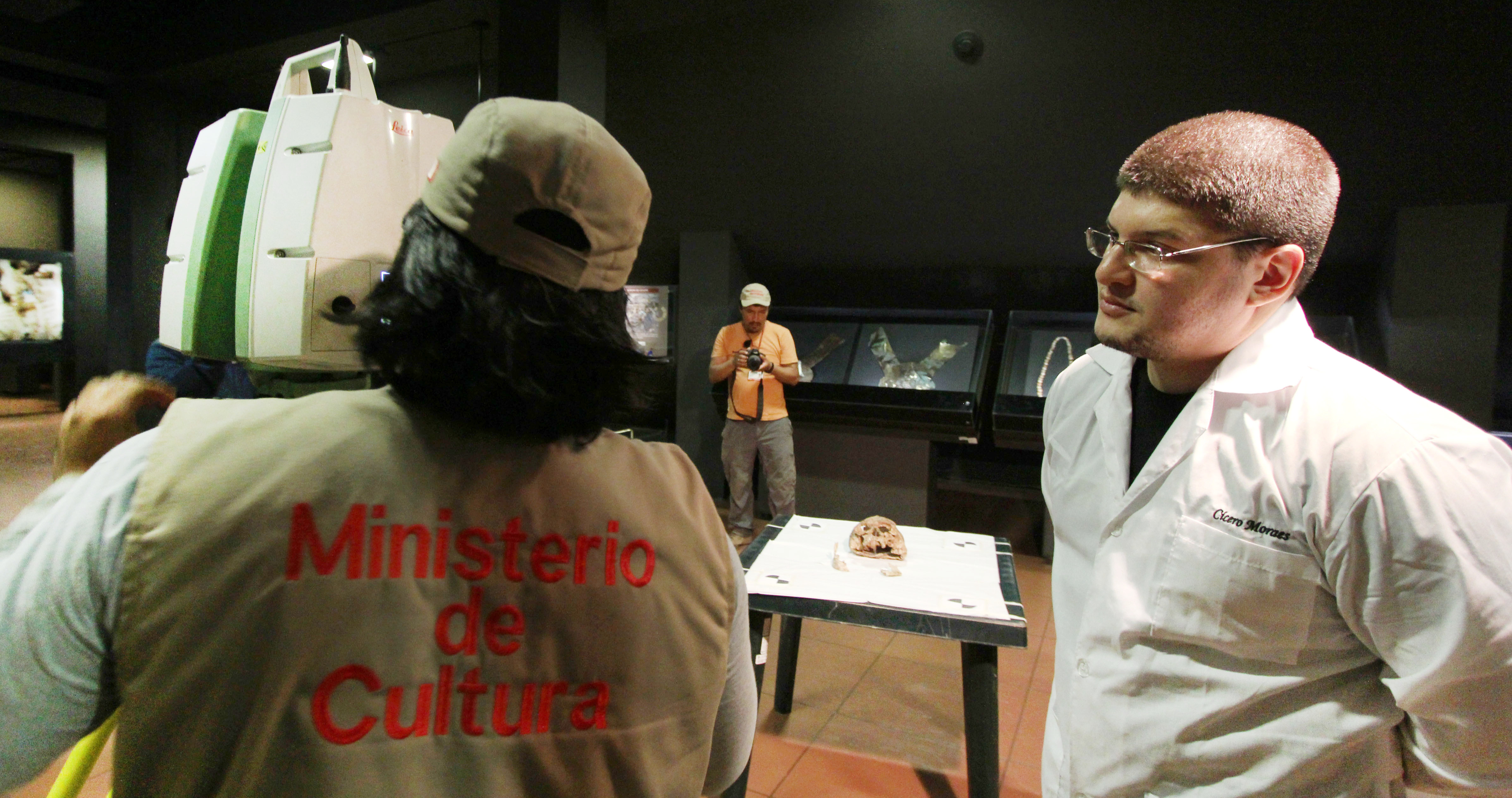
Fig. 9 Specialist from the Ministry of Culture of Peru initiating the three-dimensional digitization process of the skull of the Lord of Sipán (center), under the supervision of the author and developer C.M.
Note
A relevant factor to be highlighted in this experiment was the use of the physical skull of the Lord of Sipán as the basis for structural observations—one of the most important archaeological discoveries in the world, whose reference laser scan was executed by the Ministry of Culture of Peru (Fig. 9). This background highlights that, although OOB was officially released in 2017, its technological incubation and development process began in an earlier period. Indeed, the first trial aimed at validating the high precision of the photogrammetry pipeline consisted of the design and manufacture of a customized dental element for a specimen of Canis lupus familiaris. Based on an anatomical model digitized by photogrammetry, the resulting cobalt-chromium alloy prosthesis was subjected to physical fit tests, presenting perfect adaptation and clinical seating, which empirically proved the submillimeter accuracy of the optical reconstruction method.
Although the photogrammetry protocol had already been validated on dry skeletal structures, generating consolidated operational guidelines (https://www.ciceromoraes.com.br/doc/pt_br/OrtogOnBlender/Fotogrametria_Cranio.html), its transposition to surgical planning demanded application on living facial surfaces. This scenario imposed additional complexities, such as involuntary micro-movements of the subject during captures, variations in ambient lighting, tegumentary chromatic homogeneity in young patients, and optical reflectivity resulting from the natural oiliness of the skin. To mitigate these variables, 12 individuals of different age groups and ancestries were selected, whose faces were digitized and processed across four distinct systems. Additionally, one of the facial surfaces was captured under different illuminance conditions, generating a significant volume of raw data (https://www.ciceromoraes.com.br/doc/pt_br/OrtogOnBlender/Fotogrametria_Face_Compara.html). This database supported the development of a simplified facial digitization protocol consisting of 26 photographic takes, with an average acquisition time of 29 seconds, combined with accessible facial interventions that optimize accuracy and mitigate capture errors (https://www.ciceromoraes.com.br/doc/pt_br/OrtogOnBlender/Fotogrametria_Face.html). This methodology was extended to derived workflows aimed at the detailed reconstruction of nasal morphology [Moraes_et_al_2020_a], serving as the basis for peer-reviewed case reports published in the field of rhinoplasty—a specialty with high metric demands that employs these digital data in the manufacture of physical positioning guides to assess intraoperative fidelity in relation to the virtual plan [Sobral_et_al_2021_a]. This same principle of surface accuracy was successfully applied to the design and manufacture of customized facial prostheses [Salazar_et_al_2022_a].
Note
In the scope of computational trials, the initial experiment sought to measure the performance of the algorithm on different platforms and hardware configurations. On that occasion, the Windows environment presented a subtle time advantage compared to GNU/Linux, followed by macOS. The fastest benchmark for the add-on’s test routine was recorded on a workstation equipped with an Intel Core i9-9900K processor under Windows (7.9 s), followed by the same specification running Linux (8.0 s) [Moraes_Dornelles_Rosa_2020_a]. Alternative approaches were also tested, including the use of video sequences for three-dimensional reconstruction, which allowed the consolidation of a specific tool for dynamic facial digitization [Moraes_Dornelles_Rosa_2021b_a]. Although acquisition via static photographs remains the preferred guideline, processing video streams enabled an innovative application in the documentation and urological measurement of Peyronie’s disease, demonstrating metric equivalence when compared to traditional diagnostic methods [Nascimento_et_al_2023].
Despite the wide validation of the obtained facial geometric proportions, the process of resizing and scale calibration remained dependent on manual interventions and the technical expertise of the operator. Furthermore, there was a scarcity of scientific data regarding the pipeline’s accuracy in macro photographs of small objects. Following the evolutionary flow of the project, where preliminary chapters of the OOLM originate subsequent indexed publications, investigations focused on submillimeter precision were conducted. The first small-scale trial used a plaster dental arch, attesting to significant geometric accuracy with a distribution peak centered on the zero of the deviation histogram, confirming the viability of the technique for small bodies (https://www.ciceromoraes.com.br/doc/pt_br/OrtogOnBlender/Fotogrametria_Arcadas.html)—which in turn had already been tested and confirmed in the aforementioned case of the canine tooth printed in cobalt-chromium. Subsequently, a methodologically extreme approach compared the photogrammetry output with a native three-dimensional model perfectly free of physical errors, photographed in a virtual environment. In that study, the solution based on the OpenMVG+OpenMVS integration demonstrated greater structural compatibility with proprietary commercial solutions than competing open-source platforms, approaching the nominal reference mesh [Moraes_et_al_2022_a]. During the same period, a protocol based on ArUco-fiduciary markers was implemented to automate routines for cleanup, spatial alignment, and real-scale metric resizing, confirming purely submillimeter deviations across multiple trials [Moraes_et_al_2022b_a]. Years later, the findings of both chapters were unified and updated for the OOB XP architecture, with optimized algorithms for contemporary Python libraries. This new study stressed the system’s capabilities by evaluating the minimum and optimal number of fiduciary markers required and by comparing the level of detail of the mesh against market-leading photogrammetry software. In this evaluation, the OpenMVG+OpenMVS engine of OOB XP not only achieved technical equivalence with high-cost commercial applications, but alternated between second and third position in the global ranking of three-dimensional accuracy (volumetric and fine details) [Moraes_et_al_2025_a]. These findings were fully corroborated by an independent peer-reviewed study, which ratified the submillimeter precision of the add-on and its total viability to replace achromatic tomographic meshes in virtual surgical planning workflows [Vitorino_et_al_2026_a].
CT-Scan
One of the fundamental pillars of virtual surgical planning is computed tomography, primarily expressed through three-dimensional meshes reconstructed from DICOM standard files. The initial protocol containing best practices for radiological image acquisition was originally made available in the official OOB documentation. At the time, the document provided technical guidelines to facilitate the segmentation and separation of dental elements for subsequent replacement with high-resolution digitized arches, in addition to proposing solutions to mitigate potential metadata incompatibility issues in DICOM files (https://www.ciceromoraes.com.br/doc/pt_br/OrtogOnBlender/Tomografia.html). Within a few years of development, OOB began to offer two automated systems for 3D tomographic reconstruction: one based on the Slicer+DicomToMesh integration [Moraes_Dornelles_Rosa_2021_a] and another supported by the VTK+SimpleITK pipeline [Moraes_et_al_2021_a], in addition to enabling direct volumetric rendering on voxel data [Moraes_et_al_2021b_a].
Note
By virtue of the flexibility inherent to free and open-source software solutions, varied methodological approaches were tested in the OOB ecosystem regarding the processing of computed tomographies. Among these, the development of a script for converting video sequences into structured DICOM files for subsequent reconstruction stands out (https://www.ciceromoraes.com.br/doc/pt_br/OrtogOnBlender/VideoToDCM.html), whose objective was to transpose to the 3D environment the significant volume of linear tomographic records of biological structures publicly available on the internet. This tool was inspired by a cooperative project of one of the authors with the team at the CTI Renato Archer (a research unit subordinated to the Brazilian Ministry of Science, Technology, and Innovation), in which the video recording of the tomography of a mummy from the Egyptian-Roman period was processed for the isomorphic extraction of its skeleton and subsequent execution of a forensic facial approximation, generating a publication in an international conference [Moraes_et_al_2013_a]. The inverse workflow was also implemented in the add-on, allowing a native Blender 3D geometric mesh to be converted into a synthetic DICOM file, providing specialists with a tool to communicate with hospital systems and proprietary software dependent on this normative standard [Moraes_et_al_2021c_a] [Moraes_et_al_2021d_a].
Regarding computational performance trials, the inaugural benchmarks based on manual reconstruction revealed superior optimization in the GNU/Linux environment, followed closely by macOS and, with greater operational distance, by Windows [Moraes_Dornelles_Rosa_2020b_a]. This same efficiency landscape was recorded in the solution based on Slicer+DicomToMesh [Moraes_Dornelles_Rosa_2021_a], a scenario that was reversed only with the implementation of the VTK+SimpleITK pipeline [Moraes_et_al_2021_a], which positioned Windows (versions 8 and 10) in first place in processing speed, followed by the Linux and Mac platforms. However, under the contemporary architecture of Windows 11, the system has presented significantly longer execution times, situating itself in third place among the evaluated platforms, as detailed in the sequence.
With the popularization of deep learning models between 2023 and 2024, discussions regarding applied artificial intelligence were incorporated into the OOB user community, culminating in the initial proposal of algorithms focused on automated anatomical segmentation and the predictive localization of fiduciary landmarks [Moraes_et_al_2024_a]. This preliminary solution, however, did not obtain immediate widespread adoption by specialists, since the community demonstrated full satisfaction with the accuracy conferred by the traditional system based on scanner databases. Another limiting factor related to the requirement of specific hardware equipped with NVIDIA GPUs, given that AI segmentation routines strictly depend on the parallel CUDA architecture. It is relevant to record that the precursor experiment involving artificial intelligence in OOB dates back to 2021, with the integration interface of MeshSegNet, aimed at the automatic visual segmentation and colorization of isolated dental elements in surface meshes [Moraes_and_Dakir_2021_a]. Although promising, the tool was not absorbed by the clinical workflow of the time, returning in later iterations to act coordinately in conjunction with reconstructed meshes from computed tomographies.
During the process of porting the tools to the unified environment of OOB XP, the traditional legacy system was extensively refined. A series of more than 400 stress tests was conducted to evaluate the stability of the script against updated versions of exogenous Python libraries, succeeding in solving historical memory overflow and format incompatibility bugs. As a reflection of this engineering process, the global accuracy rate and success in tomographic reconstruction jumped from ~97% to 99% and, finally, reached the nominal mark of 100% in relation to the sample of tested examinations. It is highlighted that this sample base was mostly composed of notoriously problematic files sent by community users, containing inconsistent/incompatible library configurations, which consolidated the legacy tool as a highly resilient and reliable solution in OOB XP [Moraes_et_al_2024b_a].
Attention
Although the methodological success rate reached the nominal mark of 100% within the sample universe evaluated by the community, this index reflects the mathematical robustness of the script against known failures of DICOM files, not ensuring total infallibility against files whose raw data block is corrupted at the binary level. Under such exceptional circumstances, no computational solution will be able to restructure the original geometry. Therefore, the operator must maintain constant attention to best planning practices, performing preliminary inspections on the structural integrity of the files, verifying system security permissions, and evaluating the chronology and date difference between radiological examinations and scans, in order to avoid the manipulation of anatomical structures that have undergone bone remodeling or morphological changes resulting from intercurrent clinical procedures in the interim.
Once the traditional reconstruction system was stabilized and validated, the pipeline advanced to the native integration of segmentation routines based on artificial intelligence. The first proposed methodology utilized the DentalSegmentator framework (https://github.com/gaudot/SlicerDentalSegmentator), which performed automated separation of the calvaria, mandible, upper and lower maxillae, as well as the isolation of mental nerve canals. Operating coordinately in partnership with traditional reconstruction tools by factor, a geometric solution was developed capable of unifying and compatibilizing the spatial orientation of all resulting 3D objects in the same coordinate space. This advancement eliminated the need to computationally process the outer soft tissue mesh, since it was already isolated by the automated segmentation. A dedicated graphical interface was integrated into the add-on and subjected to a standard trial involving 151 real-case tomographies. The process recorded only two AI segmentation failures: the first associated with the presence of prostheses (geometry not mapped by the original predictive model) and the second resulting from a sample from a completely edentulous individual. This trial validated the accuracy of the system and clearly outlined its operational limits and structural reliability [Moraes_et_al_2024c_a].
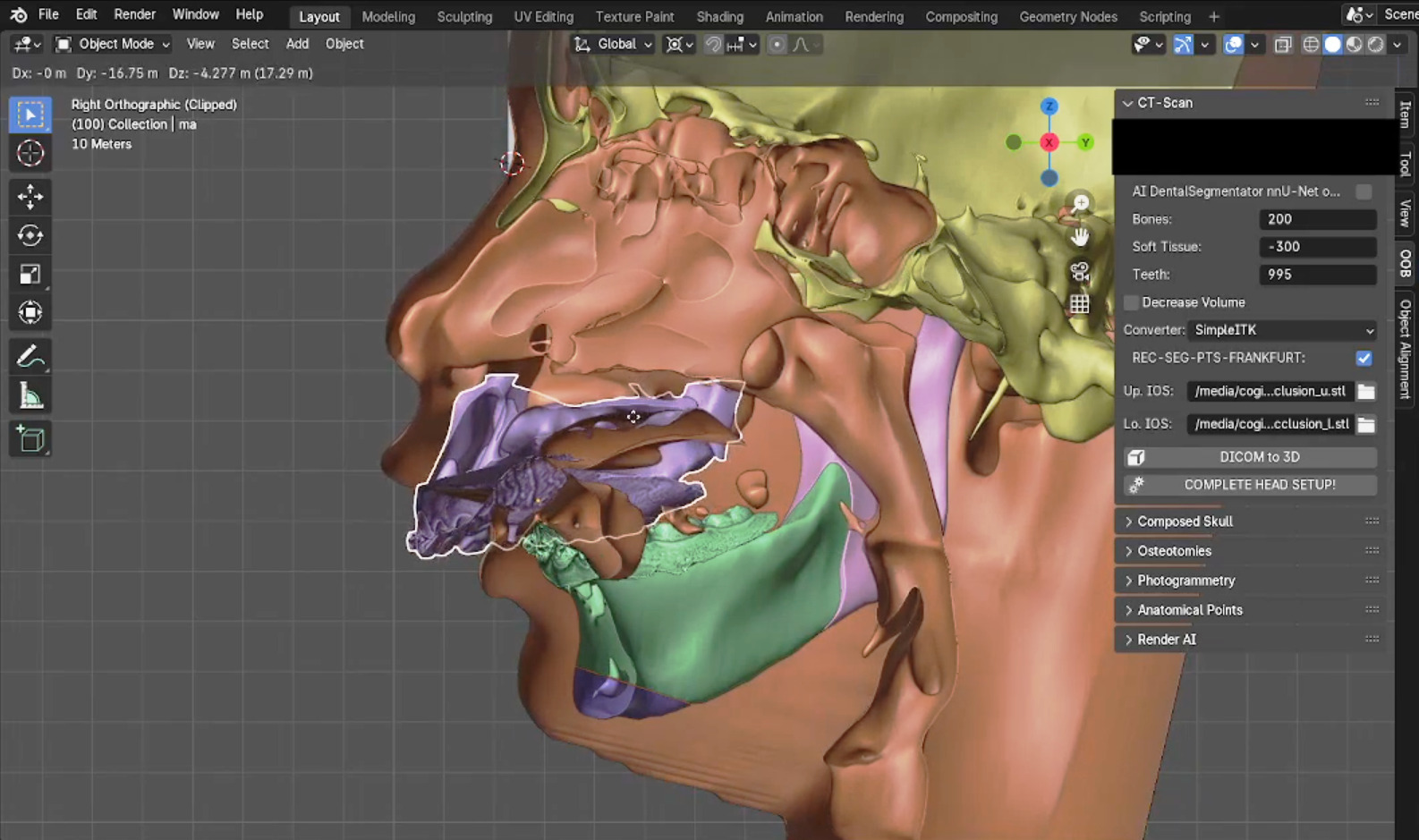
Fig. 10 Exacerbated movement test of the mandibular body after a complete automatic process. As practical evidence of the capacity and limits of this algorithmic integration, a stress test was developed simulating an exacerbated spatial movement of the mandibular body after the execution of a 100% automated workflow, encompassing the stages of tissue segmentation, alignment of intraoral scans (IOS), osteotomy tracing, and corresponding facial soft tissue deformation (https://www.youtube.com/watch?v=RRLIiqwSMKk). It is crucial to highlight that, while the computational viability and autonomous execution of the process were satisfactorily demonstrated in this controlled trial, the fully automated configuration was deliberately disabled in the source code of the final stable version of the system. This safety block was implemented to mitigate potential inconsistencies or anatomical deviations resulting from failures in algorithmic inferences, ensuring that the control of each step remains under the mandate, technical monitoring, and mandatory approval of the operating surgeon.
This methodological approach was expanded for the conception of the first Unified System for the Creation of a Composite Skull in OOB XP. The protocol demonstrated the technical feasibility of executing all preliminary stages of orthognathic surgery in a fully automated manner, extending from composite skull alignment to osteotomy simulation, although fully automated operation without human supervision is not the recommended clinical guideline. During the validation phase of this unified system, exhaustive trials were conducted in sample blocks: 115 tests dedicated to locating anatomical points and fiduciary landmarks directly on computed tomographies; 101 trials on facial meshes for determining soft tissue points; 120 evaluations of the integrated pipeline without the dental alignment stage; and 12 complete tests executed simultaneously on three distinct operating systems. This multi-distribution benchmark totaled 36 global repetitions, showing a subtle time optimization in favor of the native GNU/Linux environment compared to Windows and WSL (Windows Subsystem for Linux) [Moraes_et_al_2025b_a].
Despite the architectural advancement represented by the unified system and the evident simplification of the operational workflow when compared to the purely analog methodology, some technical aspects motivated new investigations. Among these, the fluctuating success rate in the automated alignment of high-resolution intraoral models and slight discrepancies in quality and polygonal density in meshes segmented under Windows compared to Linux stood out, which limited the total isomorphic portability of the code across platforms.
To resolve these inconsistencies, new solutions were tested, resulting in the proposal of a methodology based on the TotalSegmentator predictive model framework [Wasserthal_et_al_2023_a]. This library enabled the execution of segmentation routines in a substantially shorter window of time, adding the capacity to isolate each dental element individually. This functionality, operating in convergence with the MeshSegNet algorithms [Moraes_and_Dakir_2021_a], consolidated itself as a precise pipeline for the automatic alignment and registration of intraoral models (double-scan), presenting homogeneous and compatible results on both Linux and Windows platforms. Additionally, the system was expanded with complementary post-processing modules, now emitting automated standard USP craniometry reports and computerized cephalometric analyses of soft tissue facial proportions and boundaries.
Note
The integrated facial proportion analysis system is a component derived from the ForensicOnBlender submodule, originally developed to guide the projection of muscle and tegumentary structures in soft tissues and for the three-dimensional reconstruction of bone mass loss in archaeological calvariae. The volume of radiological data measured for the statistical validation of this predictive model was significant, beginning with the proposal of guidelines for the spatial projection of nasal morphology [Moraes_et_al_2021e_a], a study that driven the development of linear projections for predicting anatomical landmarks [Moraes_et_al_2022c_a]. Initially structured from a Brazilian population sample of mixed ancestry, the fiduciary database was expanded with the mapping of a sample of Malaysian individuals [Moraes_Abdullah_and_Abdullah_2022_a]. Both approaches were validated through an international peer-reviewed publication [Abdullah_et_al_2022_a], with the model subsequently strengthened by the incorporation of a sample of European anthropometric data [Moraes_and_Suharschi_2022_a]. This consolidated database supported the digital facial reconstruction and multidisciplinary investigation of important global historical figures, such as Ludmila of Bohemia [Moraes_et_al_2023_a] and Jan Žižka [Moraes_et_al_2024d_a] in the Czech Republic, as well as the Pharaohs Tutankhamun [Moraes_et_al_2023b_a] and Amenhotep III [Moraes_et_al_2024e_a] in Egypt. With the technical maturity of the pipeline, the facial projection and average tool began to be applied clinically in the evaluation of patients with severe structural deformities, allowing the objective measurement of the level of morphological deviation in relation to population averages. This resource was absorbed and made available directly in the unified user interface in OOB XP.
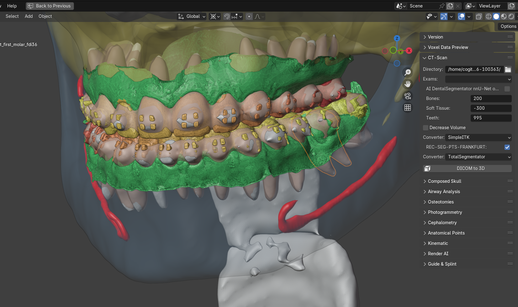
Fig. 11 Automatically generated composite skull, with IOS alignment in relation to the dental arch, with a selected segmented molar. The process was done without human interaction.
For the statistical validation and technical approval of this new unified pipeline, a series of controlled trials was conducted. One was composed of 24 pairs of IOS, and resulted in an average of 24 seconds for dental segmentation, and an average of one error per pair using MeshSegNet. Another test consisted of 23 paired segmentations of dental arches, totaling 46 processed pieces. Additionally, 23 complete craniomaxillofacial reconstructions were executed in a mirrored manner on two operating systems (Linux and Windows), totaling 46 platform consistency trials, followed by another 23 complete reconstructions using the previous method based on DentalSegmentator. Finally, aiming to assess the long-term global stability of the algorithm, the system processed an additional 37 complete reconstructions in a Linux environment, totaling 60 examinations analyzed strictly under this distribution. The benchmark results indicated that the complete tomographic processing demanded an average of 11 minutes in a GNU/Linux environment and 16 minutes under Windows. Under the perspective of algorithmic evolution, the new combined TotalSegmentator+MeshSegNet approach (11 minutes) proved to be significantly more efficient than the legacy workflow based on DentalSegmentator, which required an average of 20 minutes of processing under the same Linux platform. Regarding automation efficacy, the success rate in aligning independent arches reached 97.8% in Linux and 93.2% in Windows; when evaluated under a strict tolerance criterion focused on the examination as an indissociable unit—in which the failure of a single arch disqualified the global processing—the success index was fixed at 95.65% in the Linux environment and 86.36% in Windows, marks substantially superior to the efficiency rate recorded by the previous automated system, which had achieved only 4.35%. Even in the study with 60 cases under Linux, it maintained global accuracy precisely at 95%. These data show that the new unified composite skull import system represents a significant advance in OOB XP engineering, mitigating the time consumed in repetitive modeling tasks and allowing the specialist to concentrate efforts on the clinical and surgical analysis of the patient [Moraes_et_al_2026_a]. It is important to inform that, even if the arches are not aligned automatically, OOB was designed to allow manual corrections to be implemented by the user.
Attention
The technological advance documented in the unified system routines demonstrates the technical viability of automating the stages of segmentation, three-dimensional alignment, osteotomies, and proposal of osteotomy advancement vectors based on deviations quantified in USP craniometry and Arnett analyses (the latter in the implementation phase). However, as will be detailed below, OOB was structurally conceived as a supportive and assistive SaMD, operating under the premise of mandatory human-in-the-loop supervision. Any and all simulations executed by the software remain under the exclusive endorsement and full technical and legal responsibility of the operating surgeon. The automations and code simplifications aim solely to optimize digital workbench time so that the specialist can apply their clinical expertise to review and fine-tune the therapeutic plan. It is not recommended, expected, or endorsed by the developers that OOB, in its versions 291 or XP, be used as an autonomous decision-making or surgical planning tool.
Virtual Surgical Planning
OOB was originally conceived with the purpose of empowering specialists in the application of computer graphics to virtual surgical planning. Since its early stages, the add-on featured a structured clinical protocol, albeit with limited automated routines and a heavy reliance on the technical expertise of the operator (Orthognathic Protocol: https://www.ciceromoraes.com.br/doc/pt_br/OrtogOnBlender/Protocolo_Planejamento_Ortognatica.html). This methodology was extensively tested and peer-reviewed, with its accuracy and clinical efficacy validated through comparative analyses between pre-operative digital simulations and actual post-surgical outcomes [Cunha_et_al_2020_a] [Lobo_et_al_2022_a], as well as concurrent validation trials against high-cost commercial and proprietary software consolidated in the medical market [Lobo_et_al_2022_a]. This methodological approach demonstrated a high level of reliability and tradition, to the point of fostering a natural resistance among users to migrate to or test the new OOB XP architecture, given the high level of satisfaction and operational comfort established with the stable OOB 291 release. Diverging from the conventional flow of technological obsolescence, the developers chose to maintain active support and not discontinue the OOB 291 version, implementing continuous improvements to its tools and backporting consolidated solutions from the OOB XP environment. Rather than stagnating the project, this engineering strategy provided greater stability and safety to conduct exhaustive testing and refinement of the OOB XP architecture, making the cutting-edge version substantially more robust and predictable.
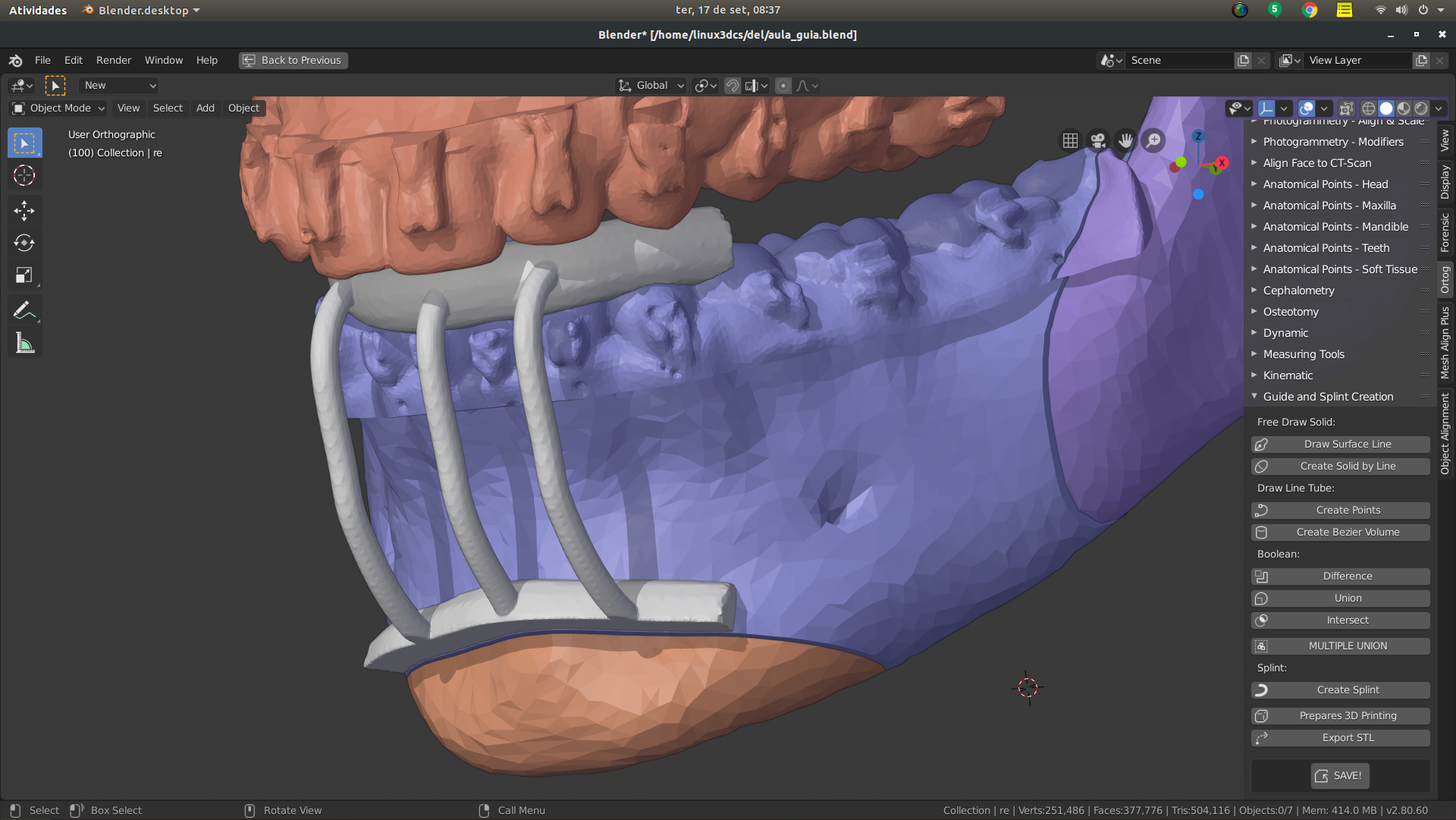
Fig. 12 Cutting guide, capture from the traditional planning protocol of OOB 291 (https://www.ciceromoraes.com.br/doc/pt_br/OrtogOnBlender/Protocolo_Planejamento_Ortognatica.html).
The scope of clinical applications linked to OOB has registered exponential growth in recent years, proving that the flexibility of open-source software, combined with the reliability of validated algorithms, provides a vast field for interventions that transcend conventional orthognathic surgery. Currently, the ecosystem encompasses consolidated workflows in subfields such as: structured rhinoplasty [Sobral_et_al_2021_a], manufacture of customized facial prostheses for oncological patient rehabilitation [Salazar_et_al_2022_a], complex urological modeling and measurement [Nascimento_et_al_2023], guided reduction of severe mandibular fractures [Facanha_25_al_2021_a], forensic facial reconstruction and approximation [Moraes_et_al_2025c_a], indirect orthodontic bonding [Gianvechio_and_Moraes_2020_a], minor dental surgical procedures (encompassing the excision of maxillary cysts [Moraes_et_al_2020b_a] and the digital management of gummy smiles [Adami_et_al_2021_a]), customized orbital reconstruction (https://ortogonline.com/doc/pt_br/OrtogOnLineMag/12/Fiocruz.html#id60), virtual planning of complex surgical flaps (https://ortogonline.com/doc/pt_br/OrtogOnLineMag/12/Fiocruz.html#id61), and determination of vectors for mandibular distraction osteogenesis [Duarte_et_al_2023_a].
A sample census conducted voluntarily with the online community of OrtogOnBlender users sought to quantify the global volume of procedures performed under the ecosystem. The statistical survey recorded a mark exceeding 2,300 virtual surgical plans completed by the network’s specialists, consolidating the tool’s practical robustness and institutional penetration within the international medical landscape.
Structural Robustness and Fiduciary Reliability
The deployment of OOB is intrinsically associated with virtual surgical planning, which demands significant accuracy of geometric measurements, given that these data will support definitive structural interventions through cutting and positioning guides manufactured by 3D printing. Under this clinical prerequisite, dimensional precision must be strictly submillimeter in all spatial coordinates.
There are records in the user community reporting historical issues with competing platforms where failures occurred due to inadvertent geometric mirroring of computed tomographies. In these scenarios, the right anatomical plane was inverted with the left (and vice versa), a technical anomaly that was frequently detected only in advanced stages of the workflow, when the physical guides had already been printed. Additionally, systemic failures can occur from corrupted DICOM files or those lacking native slice thickness metadata, which drastically alters the object’s scale or generates severely distorted polygonal meshes. Added to this is the risk of physical decalibration of 3D printers and other exogenous variables that are not linked to the modeling algorithm itself, but compromise the final result.
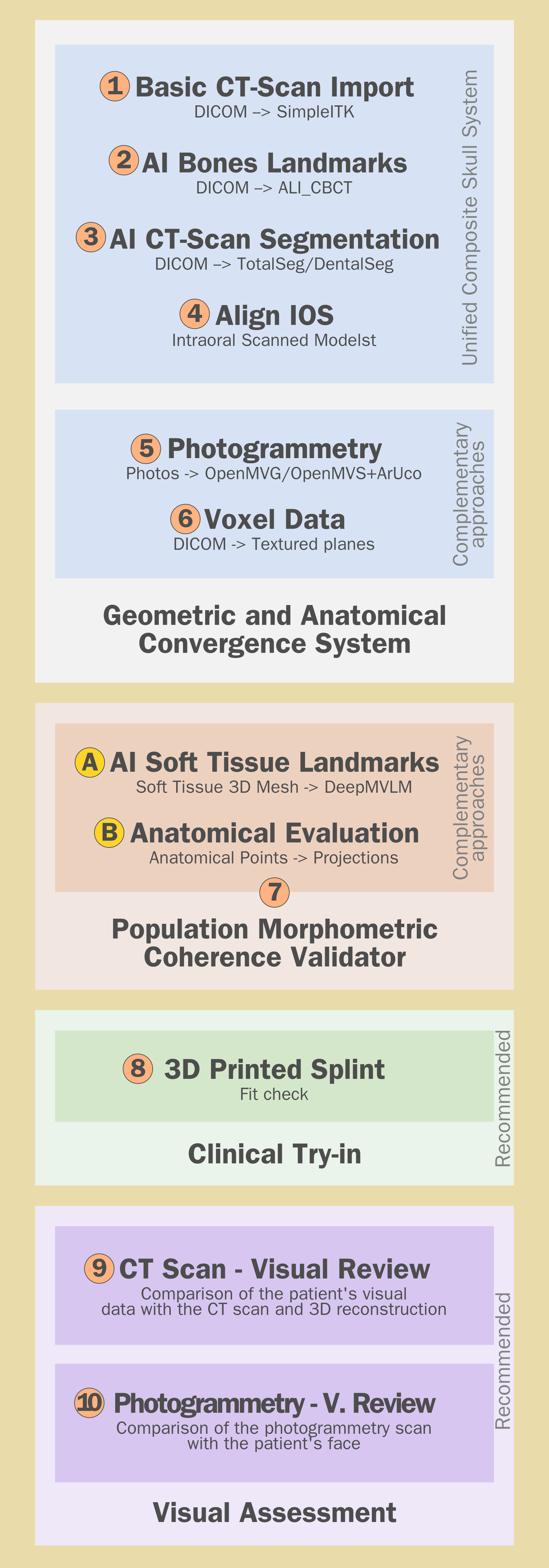
Fig. 13 Integrated system for multi-layered structural validation and checking.
The OOB ecosystem bypasses this matrix of vulnerabilities through fiduciary safety blocks which, combined with virtual planning best practices, significantly mitigate the probability of operational failures due to the high number of cross-verification steps.
As discussed preliminarily, thousands of surgical procedures have already been performed under the OOB ecosystem. In the early stages of development, reports of examinations that failed during automated reconstruction were a relatively common occurrence, prompting direct contact from users with technical support. This variability stemmed from the wide geographical dispersion of operators, who manipulated data from different manufacturers, models, and scanner calibrations, in addition to multiple intraoral scan formats.
Throughout the add-on’s maintenance cycle, these inconsistencies were systematically resolved and mitigated at the code level through automated scripts. This process highlights an artisanal refinement applied to software development, in which the algorithm was gradually polished to respond to the demands and nuances of the users’ clinical routine. As a result, the set of scripts consolidated a series of logical guidelines that ensure the stability of the planning pipeline.
Currently, the occurrence of a computed tomography configuration not mapped in the system’s native database is an extremely rare event, and the same predictability applies to the automatic screening and organization of examinations. Thus, although any digital platform is subject to latent failures, the probability of errors in tomographic reconstruction by OOB has been reduced to marginal rates.
The volumetric rendering tool (voxel data) was developed to act concurrently with the 3D mesh extraction system based on the Hounsfield scale. In comparative trials, high spatial compatibility was recorded between both methods, despite deriving from distinct mathematical pipelines—the first based on isomorphic density thresholds and the second on the projection of two-dimensional textured planes with rigid fiduciary spacing—using the same DICOM file metadata.
With the advent of segmentation tools based on deep neural networks, questions have emerged in the literature regarding the precision of these inferences when confronted with the actual anatomy expressed in the computed tomography. In light of this, the decision to preserve the traditional and legacy reconstruction engine of OOB was justified by two critical factors: first, to provide the facial soft tissue mesh without the need for additional computational processing; second, to generate reference meshes of the skull and dense cortical structures to serve as a template against the outputs generated by artificial intelligence. The logical premise dictates that if two distinct mathematical approaches generate structurally isomorphic segmentations in the same 3D space, the geometric coherence of the model is confirmed. Additionally, considering that AI algorithms often isolate complex structures in a more refined manner than pure Hounsfield thresholds, the voxel data reconstruction was used to audit these results, given that volumetric rendering preserves dense tissue nuances. This configures a triple structural validation system, converging different rendering methodologies within the same three-dimensional environment.
In the event that the tomographic file presents intrinsic structural distortions of unknown origin, the ecosystem allows the radiological data to be cross-referenced with fiduciary information from two independent optical capture methods: intraoral scanning (IOS) and facial photogrammetry. The IOS model acts as a high-fidelity fiduciary reference, since its positional precision is natively superior to that of computed tomography; therefore, the perfect and submillimeter alignment between the dental surfaces of the IOS and their tomographic counterparts attests to the dimensional integrity of the reconstructed bone structure. Any temporary orthodontic discrepancies resulting from intercurrent treatments may limit total overlap, but preserve the scale compatibility of the dental elements. Simultaneously, facial photogrammetry, originated from convergent monoscopic captures and metrically calibrated by ArUco fiduciary markers, projects the soft tissue in real scale (1:1). The successful alignment of this optical mesh with the soft tissue extracted from the tomography confirms the global consistency of the examination, immediately highlighting severe anomalies such as axis mirroring or scale distortions.
As an additional layer of protection, the add-on incorporates the Anatomical Evaluation module, which issues automated reports (via graphical interface and text files) confronting the patient’s dimensions with anthropometric limits tabulated in population studies. If the axes or proportions of the generated model extrapolate the expected standard deviations for the human species, the system alerts the operator to the inconsistency [Moraes_et_al_2026_a].
Finally, the final stage of validation is consolidated in the physical environment through the additive manufacturing of test surgical guides and splints, allowing the surgeon to perform fit tests and fiduciary seating on the patient’s dental arches prior to surgery. This procedure concurrently validates the geometric calibration of the 3D printer and the consistency of the digital mesh, linking to the mandatory visual inspection between the morphology generated on screen and the patient’s real face.
The convergence of these structured stages allows the consolidation of a multi-layered Structural Validation and Checking System (Fig. 13). Under this perspective of technical governance, OrtogOnBlender goes beyond acting as a mere modeling interface for advanced tools such as OpenMVG, OpenMVS, and TotalSegmentator, functioning as an external auditing and computational stress-testing environment under the strict supervision, endorsement, and responsibility of the operating surgeon.
Certification
The official technical training program for OrtogOnBlender is centralized and made available in a digital learning environment at: https://ortogonline.eadplataforma.app/plano/completo-semestral. The curriculum comprises an integrated package of 8 curricular modules and 155 instructional classes, complemented by asynchronous remote technical support via text and direct workstation access. Under the perspective of the technology’s international distribution, it is noteworthy that out of the 8 courses offered, 7 are taught in Portuguese and only the cutting-edge module focused on OOB XP is conducted in English; this language barrier, however, did not pose an operational limitation, as students from 38 countries successfully completed the training stages without methodological issues.
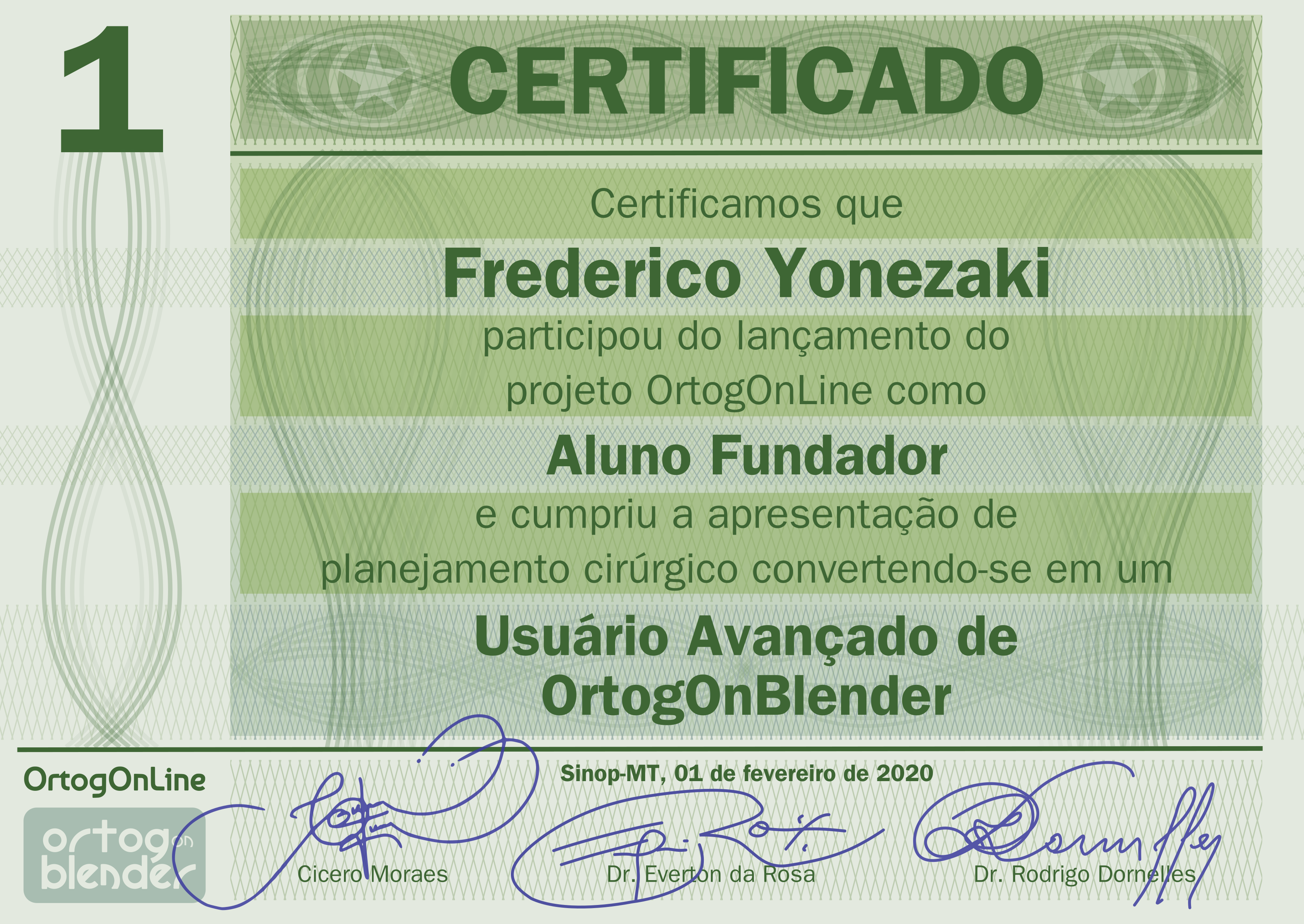
Fig. 14 Certificate of completion of the complete online training.
The granting of the official certification is not established by purely passive criteria of attendance or class participation; it strictly requires the submission and approval of a final practical virtual surgical simulation project. If the student does not present the aforementioned work, the certification document is not issued (Fig. 14).
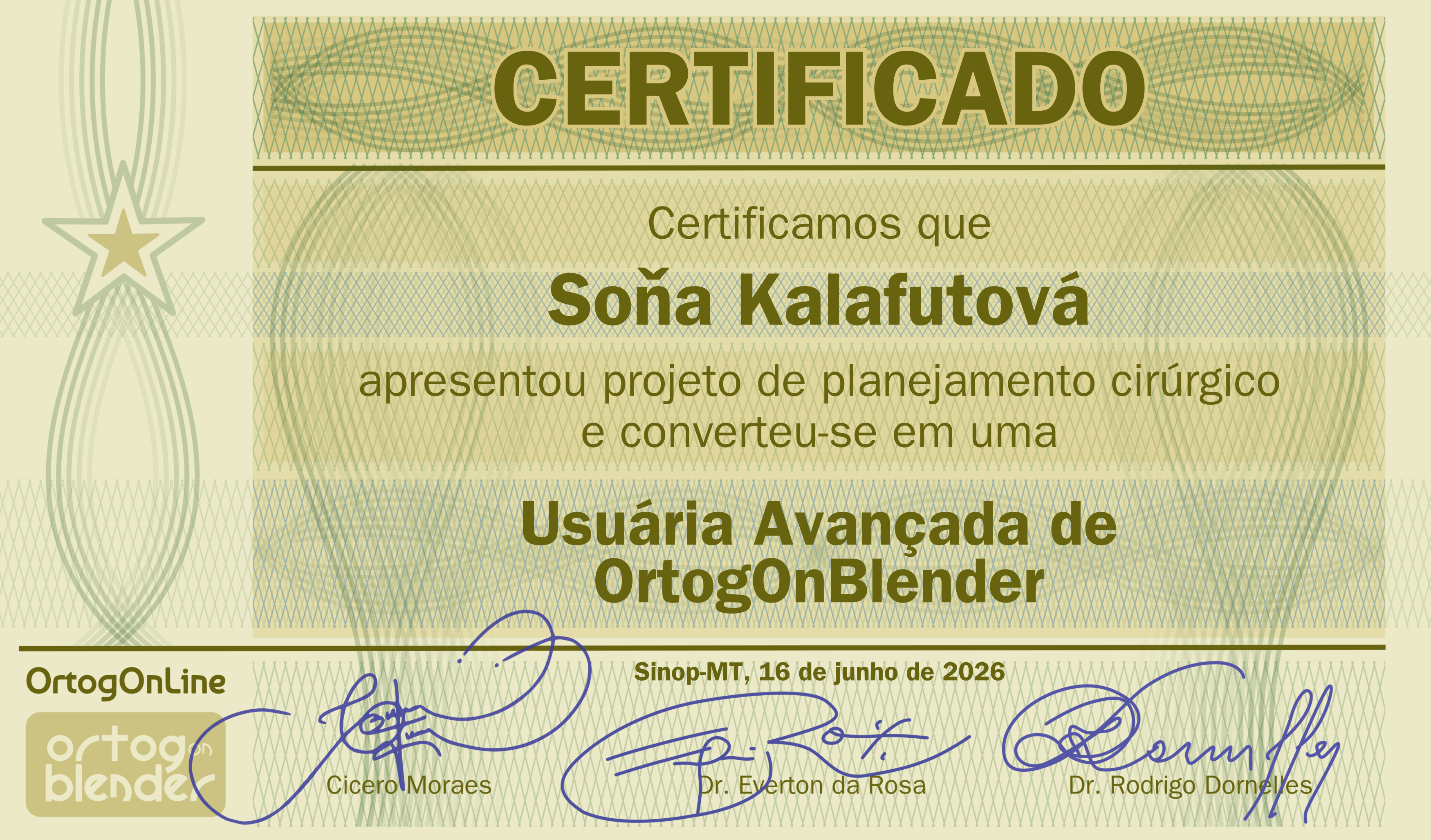
Fig. 15 Certificate of advanced proficiency.
Although the complete training requires a highly affordable financial contribution—structured to accommodate the socioeconomic reality of developing nations—it is empirically observed that a portion of potential operators are unaware of the learning platform or lack the resources for the nominal investment. In light of this, and in strict alignment with the free and open-source software philosophy that guides the project, multiple interested parties autonomously download the add-on and develop technical proficiency in a self-taught manner, supported by reading the fixed official documentation, the trials indexed in OOLM, and the audiovisual tutorials distributed on YouTube.
Whenever the successful clinical production and virtual planning of these independent users are identified by the project coordination—mostly through technical sharing on social/academic networks or through the publication of case reports in peer-reviewed journals—a certificate of advanced proficiency is honorarily awarded to these individuals, entirely free of charge, in recognition of their individual merit, self-taught effort, and reliable application of the OOB ecosystem (Fig. 15).
Conceptual Alignment with SaMD Guidelines (IMDRF) and Radical Transparency
Although the OrtogOnBlender ecosystem does not seek formal de jure regulatory certification from sanitary agencies, the technical architecture and development philosophy of the add-on establish a spontaneous (de facto) conceptual alignment with the most rigid medical software regulation frameworks in the world. Taking as reference the guidelines established by the International Medical Device Regulators Forum in the document IMDRF/SaMD WG/N10FINAL:2013 [IMDRF_2013_a], OrtogOnBlender fits precisely within the definition of Software as a Medical Device (SaMD), since it consists of software intended for medical purposes—support, investigation, and modification of human anatomy in surgical planning—that executes its functions autonomously on general-purpose computing platforms, without being embedded in or depending on the hardware of a specific physical medical device.
Additionally, under the categorization of risk model from the framework IMDRF/SaMD WG/N12FINAL:2014 [IMDRF_2014_a], OOB tools operate by crossing two fundamental axes of criticality. In the first axis, referring to the significance of the information, the software acts to Drive Clinical Management, generating three-dimensional reconstructions, craniometric reports, and virtual simulations that directly guide the decisions and subsequent interventions of specialists in the surgical routine. In the second axis, related to the state of the healthcare situation, the system operates predominantly in Serious Situations, focusing on elective reconstructive surgeries and corrections of craniomaxillofacial deformities. In this scenario, time does not configure a critical emergency of minutes, guaranteeing the surgeon a safe window to review the projections on the screen and detect fiduciary inconsistencies before the surgical act. This intersection conceptually qualifies OrtogOnBlender as a Category II (medium impact) SaMD.
To mitigate the risks inherent to this category and ensure maximum fidelity and trust in the three-dimensional measurements required in a hospital environment, the OOB lifecycle adopts the strict configuration control principle recommended by the IEC 62304:2006 standard. This is materialized in the strategic decision to freeze the operating environment for its two stable versions: OOB 291 (coupled with the immutable Blender 2.91 line) and OOB XP (frozen in Blender 4.2). This environment isolation prevents unforeseen third-party updates from corrupting the APIs or Python scripts that manage geometric calculations. This engineering posture also responds to the SOUP (Software of Unknown Provenance) control criteria of IEC 62304:2006 and risk management of ISO 14971:2007, ensuring that the integrated libraries operate under maximum predictability and metric reproducibility.
In line with the clinical evidence maturity requirements of IMDRF N12, OrtogOnBlender implements a robust system of cross-fiduciary validation composed of 10 structured and reliable steps. This multi-layered checking protocol—which involves everything from three-dimensional cross-referencing by Hounsfield factor with the generation of textured planes in Voxel Data, to metric alignment with IOS (intraoral) meshes and photogrammetry scaled by ArUco markers—acts as an active risk mitigation barrier against hidden failures common in digital environments, such as inadvertent exam mirroring or scale distortions due to the absence of native spacing data in corrupted DICOM files.
It is imperative to emphasize that, in accordance with the medical device’s Intended Use and Intended Purpose guidelines, OrtogOnBlender does not act autonomously and its outputs do not override human diagnosis. The software operates under the strict endorsement and entire responsibility of the operating surgeon. For this reason, the ecosystem’s best practices documentation emphasizes the obligation of the specialist to conduct constant visual audits, rigorously confronting the images and three-dimensional models generated on screen with the patient’s actual anatomical features and proceeding with prior physical fit tests (such as 3D printing of test surgical splints).
The robustness of this lifecycle and usability is inseparably supported by vast and detailed technical documentation. The ecosystem features fixed official documentation specifically dedicated to the frozen release version OOB 291, which has been used successfully and stably over the years by the global community, proving in clinical practice the resilience of the system and the mitigation of chronic platform operational failures. The coexistence of this consolidated historical documentation with the uninterrupted volumes of OrtogOnLineMag functions as a continuous usability engineering trail in full compliance with the IEC 62366:2007 standard. By transparently exposing internal limitations and historical algorithmic success data, the project neutralizes the “black-box” effect, providing the surgeon with the logical support necessary to exercise their role as final auditor and endorse the therapeutic plan with full legal and technical security.
The good faith of the project manifests itself in fully opening the source code, allowing the academic community and surgeons to audit every line of script in a transparent and radical manner. This extreme openness inverts traditional market logic: the security and structural validation of the software are not assured by a corporate counter stamp, but rather by the capacity for continuous verification, elimination of the black-box effect, and direct public mathematical auditing of its algorithms by any independent institution.
References
Abdullah, J. Y., Moraes, C., Saidin, M., Rajion, Z. A., Hadi, H., Shahidan, S., & Abdullah, J. M. (2022). Forensic Facial Approximation of 5000-Year-Old Female Skull from Shell Midden in Guar Kepah, Malaysia. Applied Sciences, 12(15), 7871. https://doi.org/10.3390/app12157871
Adami, C. R., Oliveira, R. R. de, Mattia, P. R. C., & Moraes, C. (2021). Manejo do Sorriso Gengival através de Enxertia com Cimento Ortopédico Planejada DigitalmenteUntitled Item. figshare. https://doi.org/10.6084/M9.FIGSHARE.16455063
Cunha, H. S., da Costa Moraes, C. A., de Faria Valle Dornelles, R., & da Rosa, E. L. S. (2020). Accuracy of three-dimensional virtual simulation of the soft tissues of the face in OrtogOnBlender for correction of class II dentofacial deformities: an uncontrolled experimental case-series study. Oral and Maxillofacial Surgery, 25(3), 319–335. https://doi.org/10.1007/s10006-020-00920-0
Dias, PEM, Moraes, CAC, Sousa JR, Beaini TL, & RFH, M. (2013). Escaneamento 3D por fotogrametria e software livre aplicado à Reconstrução Facial Forense. Unpublished. https://doi.org/10.13140/RG.2.2.30491.08489
Dias, P. E. M., Moraes, C. A. C., González, S., Alva, W. A., Rosa, E., & Bezzi, L. (2016). Comparação entre sete sistemas de fotogrametria para digitalização 3D de restos mortais esqueletizados. In Anais do XIII Congresso Brasileiro de Odontologia Legal e II Congresso Nacional de Antropologia Forense [Suplemento]. Revista Brasileira de Odontologia Legal - RBOL, 3(4), 19. https://portalabol.com.br/rbol/index.php/RBOL/issue/view/9
DORNELLES, R. de F. V., MORAES, C. A. da C., MARICEVITCH, J. P. B., & ROSA, E. L. S. da. (2018). RhinOnBlender. In Proceedings of 3DBODY.TECH 2018 - 9th International Conference and Exhibition on 3D Body Scanning and Processing Technologies, Lugano, Switzerland, 16-17 Oct. 2018 (pp. 69–72). Hometrica Consulting - Dr. Nicola D’Apuzzo. 3DBODY.TECH 2018 - 9th International Conference and Exhibition on 3D Body Scanning and Processing Technologies, Lugano, Switzerland, 16-17 Oct. 2018. https://doi.org/10.15221/18.069
Duarte, D. W., Fleith, I. J., Pinheiro, R. C., Machado, M. F., Zanin, E. M., & Collares, M. V. M. (2023). Mandibular morphology and distraction osteogenesis vectors in patients with Robin sequence. In International Journal of Oral and Maxillofacial Surgery (Vol. 52, Issue 4, pp. 442–450). Elsevier BV. https://doi.org/10.1016/j.ijom.2022.07.007
Façanha de Carvalho, E., Alkmin Paiva, G. L., Yonezaki, F., & Machado, G. G. (2021). Computer-Aided Surgical Simulation in Severe Atrophic Mandibular Fractures: A New Method for Guided Reduction and Temporary Stabilization Before Fixation. In Journal of Oral and Maxillofacial Surgery (Vol. 79, Issue 4, p. 892.e1-892.e7). Elsevier BV. https://doi.org/10.1016/j.joms.2020.11.011
Falih, M. R., Abed, F. M., Salazar-Gamarra, R., & Dib, L. L. (2021). Efficiency of +IDonBlender Photogrammetric Tool in Facial Prosthetics Rehabilitation – An Evaluation Study. In Karbala International Journal of Modern Science (Vol. 7, Issue 4). University of Kerbala - KIJOMS. https://doi.org/10.33640/2405-609x.3167
Gianvechio, S. L. R., & Moraes, C. (2020). Guia de Colagem Ortodôntica Indireta com o OrtogOnBlender. figshare. https://doi.org/10.6084/M9.FIGSHARE.12996908
International Medical Device Regulators Forum. (2013). Software as a Medical Device (SaMD): Key Definitions (imdrf-tech-131209-samd-key-definitions-140901_3.pdf). IMDRF SaMD Working Group.
International Medical Device Regulators Forum. (2014). “Software as a Medical Device”: Possible Framework for Risk Categorization and Corresponding Considerations (imdrf-tech-140918-samd-framework-risk-categorization-141013_3.pdf). IMDRF SaMD Working Group.
Instituto Nacional da Propriedade Industrial. (2026). Programas de computador – RPI 2892 de 09 de junho de 2026. https://revistas.inpi.gov.br/pdf/Programa_de_computador2892.pdf
Lima, G. S., Barbosa, M. D., Everton Luís Santos Da Rosa, Cicero Moraes, Souza, G. A., & Freitas, P. H. L. D. (2018). ESTUDO COMPARATIVO DA OBTENÇÃO DE FOTOS PARA A FOTOGRAMETRIA NO PLANEJAMENTO DE CIRURGIAS ORTOGNÁTICAS. Unpublished. https://doi.org/10.13140/RG.2.2.10893.64483
Lobo, F., Filho, L. I., Sigua-Rodriguez, E. A., da Silva, B. G., Tolentino, E. S., Borges, Y. M., da Silva, M. C., Tonin, R. H., & Iwaki, L. C. V. (2022). Evaluation of ortogonblender software bone movement tools in bimaxillary orthognatic surgeries performed in dolphin software. Journal of stomatology, oral and maxillofacial surgery, 123(4), 417–421. https://doi.org/10.1016/j.jormas.2021.10.001
Moraes, C., Abdullah, J. Y., & Abdullah, J. M. (2022). Mensuração de Dados Faciais Ortográficos em Malaios e Comparação com Brasileiros. figshare. https://doi.org/10.6084/M9.FIGSHARE.20000720
Moraes, C., & Dakir, I. (2021). Segmentação Automática dos Dentes com o MeshSegNet e o OrtogOnBlender. figshare. https://doi.org/10.6084/M9.FIGSHARE.14850105
Moraes, C., & Suharschi, I. (2022). Mensuração de Dados Faciais Ortográficos em Moldavos e Comparação com Outras Populações. figshare. https://doi.org/10.6084/M9.FIGSHARE.20089754
Moraes, C., Dornelles, R., & Rosa, E. da. (2020). Fotogrametria 3D - Desempenho nos Sistemas Operacionais: Windows, Linux e Mac OS X. figshare. https://doi.org/10.6084/M9.FIGSHARE.13456442
Moraes, C., Dornelles, R., & Rosa, E. da. (2020). Reconstrução de Tomografias nos Sistemas Operacionais: Windows, Linux e Mac OS X. figshare. https://doi.org/10.6084/M9.FIGSHARE.13480326
da Costa Moraes, C., Amorim, P., de Moraes, T., Ruppert, G., da Silva, J., & Santos, M. (2013). Forensic 3D facial approximation from a CT scan video of a mummified Egyptian-Roman child. In High Value Manufacturing: Advanced Research in Virtual and Rapid Prototyping (pp. 585–590). CRC Press. https://doi.org/10.1201/b15961-107
Moraes, C., Sobral, D., Duarte, D. W., Cavalcanti, G. Z., Salazar-Gamarra, R., & Dornelles, R. (2020). Protocolo Complementar para Melhor Resolução do Nariz em Fotogrametria 3D. figshare. https://doi.org/10.6084/M9.FIGSHARE.13010300
Moraes, C., Macedo, R., Alonson, G., Shiguihara, C. A. A., & Rosa, E. da. (2020). Confeccionando um Guia para Retirada de Cisto Dentário com o OrtogOnBlender. figshare. https://doi.org/10.6084/M9.FIGSHARE.13256816
Moraes, C., Dornelles, R., & Rosa, E. da. (2021). Sistema de Reconstrução de Tomografia Computadorizada Baseado no Slicer 3D e no DicomToMesh. figshare. https://doi.org/10.6084/M9.FIGSHARE.13513890
Moraes, C., Dornelles, R., & Rosa, E. da. (2021). Digitalização 3D de Faces a partir de Vídeos. figshare. https://doi.org/10.6084/M9.FIGSHARE.14075456
Moraes, C., Dakir, I., Dornelles, R., & Rosa, E. da. (2021). Reconstrução de Tomografias com o VTK Python, o SimpleITK e o Multiprocessing. figshare. https://doi.org/10.6084/M9.FIGSHARE.14370902
Moraes, C., Graf, M., Dornelles, R., & Rosa, E. da. (2021). Reconstrução de Voxel Data no OrtogOnBlender. figshare. https://doi.org/10.6084/M9.FIGSHARE.13670134
Moraes, C., Dornelles, R., Rosa, E. da, Abdullah, J. Y., & Abdullah, J. M. (2021). Conversão de Malhas 3D em Arquivos DICOM com o OrtogOnBlender. figshare. https://doi.org/10.6084/M9.FIGSHARE.14125706
Moraes, C., Dornelles, R., & Rosa, E. da. (2021). Melhoramentos na Conversão de Objetos 3D em Arquivos DICOM no OrtogOnBlender. figshare. https://doi.org/10.6084/M9.FIGSHARE.14607900
Moraes, C., Sobral, D. S., Mamede, A., & Beaini, T. L. (2021). Sistema Complementar de Projeção Nasal em Reconstruções/Aproximações Faciais Forenses. figshare. https://doi.org/10.6084/M9.FIGSHARE.17209379
Moraes, C., Bezzi, L., Bezzi, A., Šindelář, J., Rosa, E. da, & Dornelles, R. (2022). Modelo 3D vs Fotogrametria por Imagens e Vídeo. figshare. https://doi.org/10.6084/M9.FIGSHARE.20633262
Moraes, C., Kimura, R. N., Bezzi, L., & Bezzi, A. (2022). Fotogrametria com Limpeza de Pontos, Alinhamento e Redimensionamento Automáticos no OrtogOnBlender. figshare. https://doi.org/10.6084/M9.FIGSHARE.19450283
Moraes, C., Gravalos, R., Machado, C. R., CHILVARQUER, I., Curi, J., & Beaini, T. L. (2022). Investigação de Preditores Anatômicos para o Posicionamento dos Globos Oculares, Asas Nasais, Projeção dos Lábios e Outros a partir da Estrutura do Crânio. figshare. https://doi.org/10.6084/M9.FIGSHARE.19686294
Moraes, C., Šindelář, J., Galassi, F. M., & Beaini, T. L. (2023). Ludmila of Bohemia (ca. 860–921): Multidisciplinary considerations on her facial approximation. Digital Applications in Archaeology and Cultural Heritage, 29, e00263. https://doi.org/10.1016/j.daach.2023.e00263
Moraes, C., Habicht, M. E., Galassi, F. M., Varotto, E., & Beaini, T. (2023). Pharaoh Tutankhamun: a novel 3D digital facial approximation. Italian Journal of Anatomy and Embryology, 127(1), 13–22. https://doi.org/10.36253/ijae-14514
Moraes, C., Schreiner, T., Startek, B., & Beaini, T. L. (2024). Alinhamento em Frankfurt, Colocação de Pontos Anatômicos e Segmentação do Crânio por Inteligência Artificial no OrtogOnBlender. doi: 10.6084/m9.figshare.25140020
Moraes, C., Dakir, I., Startek, B., Dornelles, R., & Rosa, E. da. (2024). Ferramenta de Importação de Tomografia Computadorizada no OrtogOnBlender XP. figshare. https://doi.org/10.6084/M9.FIGSHARE.27648876
Moraes, C., Dakir, I., Startek, B., Dornelles, R., & Rosa, E. da. (2024). Segmentação de tomografias computadorizadas por ia no ortogonblender xp. figshare. https://doi.org/10.6084/M9.FIGSHARE.27761970
Moraes, C., Abdullah, J. Y., Šindelář, J., Šindelář, M., Thomová, Z., Smrčka, J., Vaccarezza, M., Beaini, T., & Galassi, F. M. (2024). The Facial Approximation of the Skull Attributed to Jan Žižka (ca. AD 1360–1424). Heritage, 8(1), 7. https://doi.org/10.3390/heritage8010007
Moraes, C., Habicht, M. E., Artico, M., Forte, F., Varotto, E., & Galassi, F. M. (2024). The mummy of <scp>Pharaoh Amenhotep III</scp> (reigned ca. 1388–1351 <scp>BC</scp>) and its facial approximation: An anatomical approach. Clinical Anatomy, 38(2), 211–215. https://doi.org/10.1002/ca.24251
Moraes, C., Moulon, P., Bezzi, A., Moura, L., Rosa, E., Dornelles, R., Bezzi, A. (2025). Photogrammetry in OrtogOnBlender XP: The Story of an (R)Evolution. figshare. https://doi.org/10.6084/M9.FIGSHARE.30656390
Moraes, C., Startek, B., Dakir, I., Schreiner, T., Dornelles, R., & Rosa, E. da. (2025). Sistema Unificado para a Criação de um Crânio Composto no OrtogOnBlender XP. figshare. https://doi.org/10.6084/M9.FIGSHARE.28135760
Moraes, C., Abdullah, J. Y., Šindelář, J., Šindelář, M., Kroupa, P., Frolík, J., Galassi, F. M., & Beaini, T. (2025). Forensic Facial Approximation of the Skull Attributed to Wenceslas of Bohemia (ca. 907–935). Heritage, 8(6), 221. https://doi.org/10.3390/heritage8060221
Moraes, C., Gravalos, R., Tadeu Ferreira Sanchis, W., Dornelles, R., & Rosa, E. da. (2026). Expansion and Automation of the Unified Import System for Composite Skulls in OrtogOnBlender XP. figshare. https://doi.org/10.6084/M9.FIGSHARE.32899760
Pierre Moulon, Alessandro Bezzi. Python Photogrammetry Toolbox: A free solution for Three-Dimensional Documentation. ArcheoFoss, Jun 2011, Napoli, Italy. pp.1-12. ⟨hal-00834940⟩
Nascimento, B. C. G., Moraes, C. A. D. C., Neto, R. P., Rocha, B. A., Miranda, E. D. P., Bessa, J. D., Nahas, W. C., Hallak, J., Mulhall, J. P., & Gomes, C. M. (2023). (167) 3D Penile Reconstruction Imaging in Complex Peyronie’s Disease (PD): Proof of Concept Study. The Journal of Sexual Medicine, 20(Supplement_1). https://doi.org/10.1093/jsxmed/qdad060.162
Pierri, R. A. G., Nogueira, L. C., Balan, I., Iwaki, L. C. V., & Filho, L. I. (2019). Bimaxillary orthognatic surgery planned with the software blender, through the addon ortogonblender. International Journal of Oral and Maxillofacial Surgery, 48, 254. https://doi.org/10.1016/j.ijom.2019.03.776
Rabello, R., Rabello, M., Fecchio R., S., Moraes, C., P.E.M., D., Palma, B., & Davanco, R. R. (2016). Utilização de Tecnologia 3D Para Reconstituição Protética de Caparaça de Jabuti (Chelonoidis carbonaria) - Relato de Caso. Unpublished. https://doi.org/10.13140/RG.2.2.34596.80001
Salazar-Gamarra, R., Cárdenas-Bocanegra, A., Masch, U., Da Costa Moraes, C. A., Seelaus, R., Lopes Da Silva, J. V., & Lauria Dib, L. (2022). Color translation from monoscopic photogrammetry +ID Methodology into a Polyjet final 3D printed facial prosthesis. In F1000Research (Vol. 11, p. 582). F1000 Research Ltd. https://doi.org/10.12688/f1000research.111196.1
Sobral, D. S., Duarte, D. W., Dornelles, R. F. V., & Moraes, C. A. C. (2021). 3D Virtual Planning for Rhinoplasty Using a Free Add-On for Open-Source Software. In Aesthetic Surgery Journal (Vol. 41, Issue 8, pp. NP1024–NP1032). Oxford University Press (OUP). https://doi.org/10.1093/asj/sjab085
Souza, M. M., Edimar Antônio Nogueira Mota, Cicero Moraes, Everton Luis Santos Da Rosa, Souza, G. A., & Freitas, P. H. L. D. (2018). ORTOGONBLENDER: A UTILIZAÇÃO DE SOFTWARE LIVRE PARA O PLANEJAMENTO VIRTUAL DE CIRURGIAS ORTOGNÁTICAS. Unpublished. https://doi.org/10.13140/RG.2.2.24315.41763
Shibasaki, W., Loiola, M., Moraes, C., Lima, L., Vieira, M. C. S., Gandini Junior, L. G., & Cotrim-Ferreira, F. (2018). Código aberto na Ortodontia - era só o que me faltava! OrtodontiaSPO, 51(5), 578–584.
Vitorino, N. de S., Dias, F. J. de N., & Daruge, R. J. (2026). Photorealistic 3D Facial Meshes Obtained by Low-Cost Photogrammetry: Are They Precise Enough to Replace Colorless CT-scan Meshes in Virtual Surgical Planning? Brazilian Archives of Biology and Technology, 69. https://doi.org/10.1590/1678-4324-2026250384
Wasserthal, J., Breit, H.-C., Meyer, M. T., Pradella, M., Hinck, D., Sauter, A. W., Heye, T., Boll, D. T., Cyriac, J., Yang, S., Bach, M., & Segeroth, M. (2023). TotalSegmentator: Robust Segmentation of 104 Anatomic Structures in CT Images. Radiology: Artificial Intelligence, 5(5). https://doi.org/10.1148/ryai.230024