3D Digital Analysis of the Anatoli Bugorski Case (1978)
Attention
This material is licensed under the following Creative Commons license: Attribution 4.0 International (CC BY 4.0).
Abstract
This study presents a three-dimensional digital analysis of the Anatoli Bugorski case, a Soviet physicist struck by a high-energy proton beam in 1978 at the U-70 synchrotron in Protvino. The accident, which exposed Bugorski to lethal radiation doses (200,000 to 300,000 rads), is reconstructed using virtual donor tomographies and tools such as the OrtogOnBlender XP add-on. The 3D reconstruction maps the beam’s trajectory through the head, from the occipital region to the left nostril, analyzing anatomical impacts in areas like the temporal lobe and temporal bone. Despite the scarcity of primary data, the results are consistent with historical medical descriptions, highlighting injuries that explain symptoms such as hypoesthesia, hearing loss, and epilepsy. The study explores the feasibility of anatomical reconstructions based on public information, offering insights into radiation effects and the brain plasticity that enabled Bugorski’s partial recovery. Beyond its scientific value, the work serves an educational purpose, providing teaching material for 3D modeling techniques in anatomy and radiobiology. Limitations include reliance on low-resolution photographs and the absence of original documentation, introducing a margin of error. Thus, the study does not replace primary data but contributes to elucidating this rare case and inspiring future research.
Keywords: Anatoli Bugorski, U-70 Synchrotron, Anatomical Reconstruction, Brain Injury, Radiation Effects, 3D Modeling, Neuroplasticity
Introduction
Soviet Context
The Union of Soviet Socialist Republics (USSR) relied on centralized planning that encompassed everything from the economy to structural organization, including the creation of cities with specific purposes. Among them, the atomgrads, or atomic cities, such as Prypiat, near the Chernobyl nuclear power plant, gained worldwide notoriety due to the 1986 nuclear accident. In addition to the atomgrads, the USSR developed around 60 scientific cities, known as naukograds, starting in the late 1940s, aimed at advancing research in strategic areas such as nuclear physics and cutting-edge technology during the Cold War.
One of these naukograds, Dubna, remains notable to this day for its scientific contributions, such as the isolation of elements 102, 103, 104, 105 (dubnium, named after the city), and 114 in 1999 [Gessen_1997_a]. Another prominent city was Protvino, which housed the largest particle accelerator of its time [Arguments_and_Facts_2020_a], the U-70 synchrotron. With a 1.5 km vacuum tube, the U-70 generated an energy of 70 billion electron-volts, a significant milestone in particle physics [Izvestia_1998_a].
The Anatoli Bugorski Accident
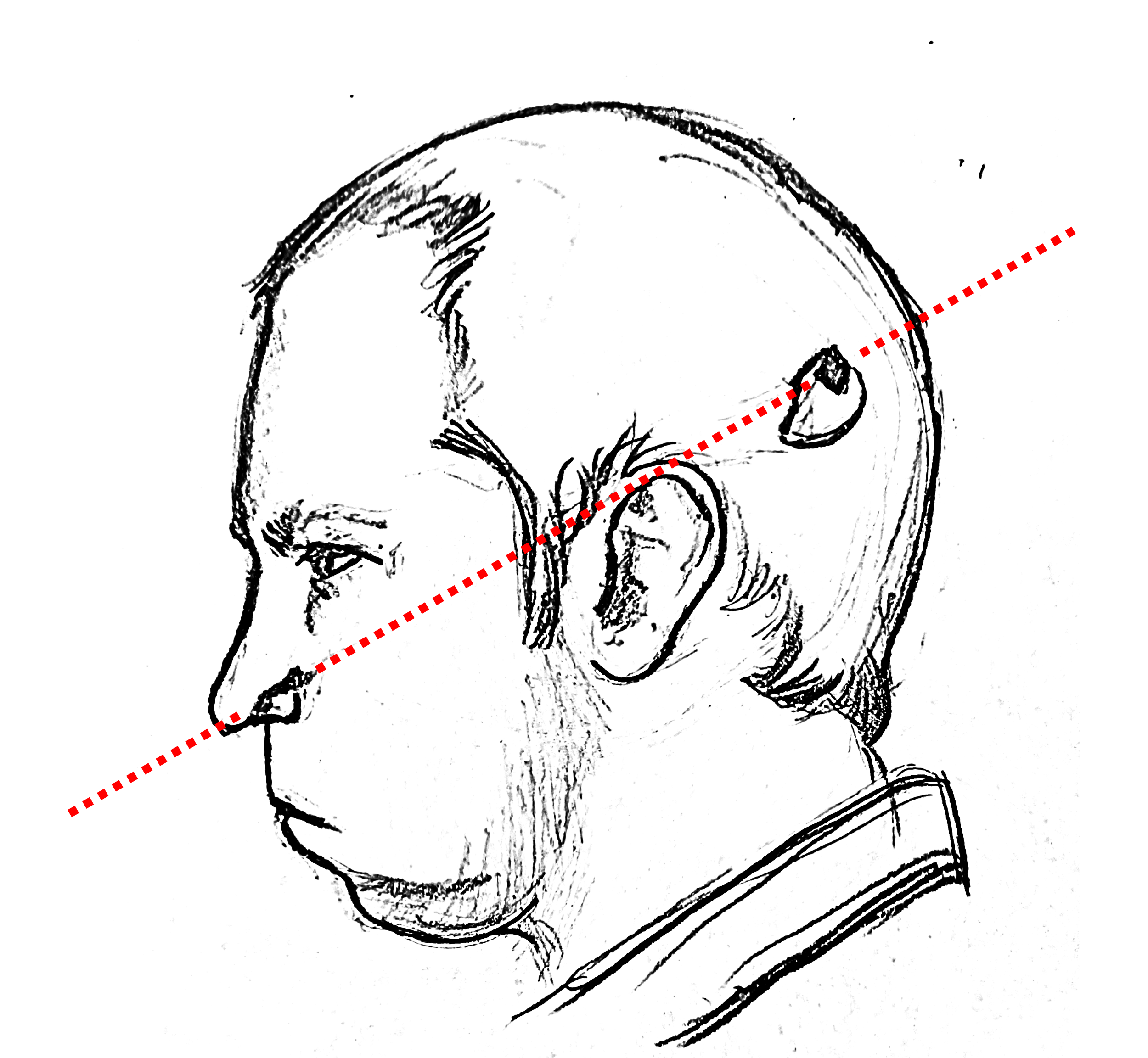
Fig. 1 Author’s drawing (C.M.) based on a photograph taken three months after the accident [Izvestia_1998_a]. The red dotted line represents the trajectory of the proton beam, which entered through the left occipital region and exited beside the left nostril.
On June 3, 1978, physicist Anatoli Bugorski, then 36 years old and a researcher at the Institute of High Energy Physics in Protvino, was called to fix a malfunction in the U-70’s detection system. Due to an operational error, the safety light indicating the operation of the proton beams was burnt out, and the access door, which should have been locked, was left open after a previous experiment. Upon entering the accelerator, Bugorski was struck by a high-energy proton beam, with a 2x3 mm cross-section, which passed through his head from the left occipital region to the left nostril (Fig. 1), exposing him to a radiation dose of 200,000 rads at entry and 300,000 rads at exit due to dispersion [Izvestia_1998_a]. For comparison, doses of 500 to 600 rads are already lethal to humans [Gessen_1997_a].
According to Bugorski, the beam’s impact produced an extremely intense flash, described as “brighter than a thousand suns” [Gessen_1997_a]. Despite the incident, he completed his task and recorded the visit in the activity log [Izvestia_1998_a]. That night, Bugorski noticed significant swelling on the left side of his face and, the next day, sought medical attention. Initially, doctors doubted his account, but the visible symptoms led to his transfer to Hospital No. 6 in Moscow, specialized in radiation injuries [Arguments_and_Facts_2020_a].
Recovery and Aftereffects
Despite pessimistic prognoses, Bugorski survived, attributing his recovery to good health and regular sports practice [Arguments_and_Facts_2020_a]. Details about the treatment are scarce due to the USSR’s secrecy regarding accidents involving high technology, but Hospital No. 6 was known for its expertise in radiation cases, which became widely recognized after the Chernobyl disaster. A year and a half after the accident, Bugorski returned to work [Izvestia_1998_a].
However, the physicist faced permanent aftereffects: he lost hearing in his left ear, experiencing only a constant buzzing; he suffered epileptic seizures with loss of consciousness; and, although showing no evident intellectual impairment, he experienced significant mental fatigue. Despite these challenges, Bugorski completed his kandidat nauk dissertation (equivalent to a Western doctorate), but exhaustion and health issues prevented him from pursuing the doktor nauk title, which would have required additional years of research [Izvestia_1998_a].
Long-Term Impact
Due to the lack of specific public policies for accidents like his, Bugorski only received state support after the Chernobyl disaster, being included in a program for radiation victims. After the dissolution of the USSR, he faced difficulties in covering treatment costs [Gessen_1997_a]. Nevertheless, he remained active at the Institute of Physics until at least 2020, at the age of 77 [Arguments_and_Facts_2020_a]. His family continues to live in Protvino, where they enjoy local prestige, as evidenced by a 2024 news report about his granddaughter’s marriage proposal during an election session in the city [Msk1_ru_2024_a].
Study Motivation
The accident involving Anatoli Bugorski, which occurred in 1978 at the U-70 synchrotron in Protvino, is a case of significant public interest, widely reported in news outlets and online media, but lacking detailed academic analyses. The primary reliable sources available are two articles published in the 1990s [Gessen_1997_a] [Izvestia_1998_a], accompanied by a limited number of reference images, mostly low-resolution photographs. Given this scarcity of scientific studies, this work aims to reconstruct the accident through a 3D digital anatomical analysis, seeking to detail the regions affected by the proton beam and verify the consistency between the injury, the immediate symptoms, and the reported aftereffects.
In addition to contributing to the understanding of the case, this study has an educational purpose: to develop teaching material for anatomical analysis techniques. Using data extracted from news reports, books, online media, and academic journals, the work tests the feasibility of reconstructing complex injuries based on publicly available information, employing 3D modeling tools to map the beam’s trajectory and its anatomical impacts. The analysis also aims to clarify how factors such as the beam’s energy (200,000 to 300,000 rads) and radiation dispersion influenced the observed damage, providing insights for students and professionals in the fields of medical physics and radiobiology.
Material and Methods
Software
The work described in this document was carried out using the OrtogOnBlender XP (https://www.ciceromoraes.com.br/doc/pt_br/OrtogOnBlender/index.html) addon, an extension developed by one of the authors (C.M.) to enable Blender to perform tasks not available in the native version, such as importing tomographies, either as voxel data [Moraes_et_al_2021_a] or as 3D meshes [Moraes_et_al_2025_a].
References Consulted
Regarding historical aspects and medical evaluations, two documents were extensively consulted due to their relevance and frequent citation in derivative news outlets. The first is the renowned Wired article, The future ruins of the nuclear age, authored by [Gessen_1997_a], which, in addition to addressing the accident, contextualizes the scientific cities project (naukograds). The article also highlights Bugorski’s interest in making himself available to Western scientists, considering himself, in his words, a valuable study subject [Gessen_1997_a]. The second material, equally significant and containing valuable images for this study, is the article by [Izvestia_1998_a], titled Anatoli Bugorski’s Personal Chernobyl, written at the request of the interviewee, which provides crucial information about the medical evaluation.
For the consultation of anatomical data and naming, the Human Anatomy Atlas by [Tank_and_Gest_2009_a] was consulted.
The analysis of the case involved a series of complex events and approaches, requiring the review of a considerable number of articles. After a selection process focused on the present case, only a portion was included in the references. The comprehensive work by [Price_20212_a], titled A review and synthesis of the first 20 years of PET and fMRI studies of heard speech, spoken language and reading, provided a detailed insight into the brain region primarily affected by the 1978 accident, consolidating 20 years of studies in an extensive document that also referenced previously consulted articles, which is why the author chose to omit them in its citation. Another relevant study was [Nieberlein_et_al_2023_a], titled Reorganization and Plasticity of the Language Network in Patients with Cerebral Gliomas, which addresses brain reorganization and the factors explaining the high success rate in cases similar to the present one. Regarding hypoesthesia and paresis, the studies by [Lee_et_al_2020_a] and [Devoti_et_al_2021_a] explored lesions in the infraorbital foramen region, while [Kurihara_et_al_2020_a] provided a wide range of explanations about traumas in the temporal bone region, complementing the discussion on loss of sensitivity and movement. Still addressing the effects of lesions in specific brain regions, the work by [Nayak_and_Bandyopadhyay_2023_a] analyzes epilepsy cases associated with activity in the temporal lobe. Finally, concerning post-traumatic fatigue in the brain region, [Wright_et_al_2023_a] presents an approach focused on patients’ experiences and practical outcomes following treatment.
Initial Analysis of the Injury
There are not many references regarding the injury suffered by Bugorski. The most widely disseminated images related to the case are those published in a 1998 newspaper article [Izvestia_1998_a], which include a lateral profile photo of the face (https://img0.liveinternet.ru/images/attach/c/1//62/450/62450161_2p5month.JPG) and an image purportedly showing the radiograph with the proton beam trajectory (https://img1.liveinternet.ru/images/attach/c/1//62/450/62450110_Trassa.jpg).
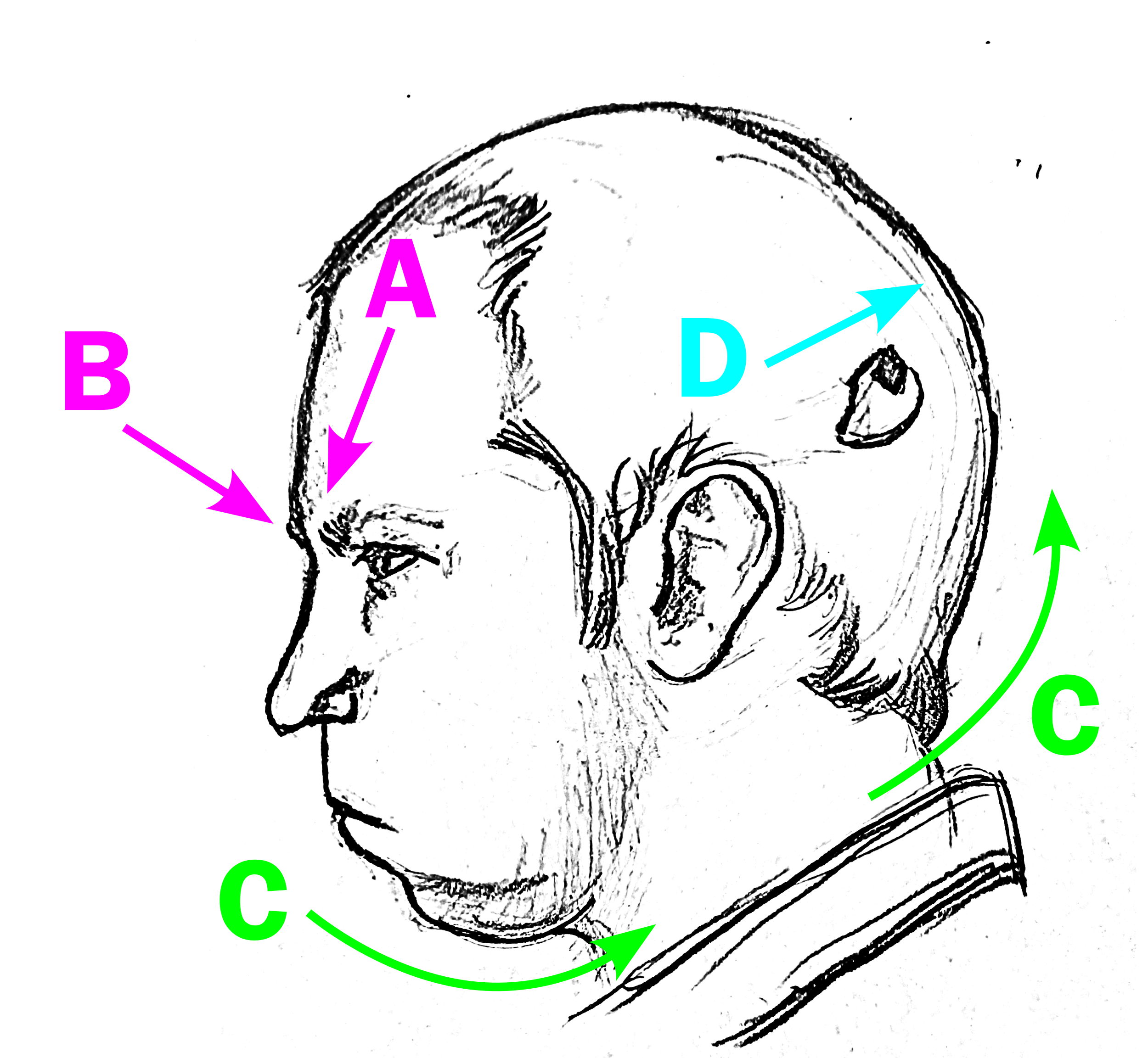
Fig. 2 Illustrative diagram regarding the slight head rotation relative to the lateral view. The use of original material respects the lack of licensing for the original photo. For further details, refer to the provided link for the photograph of Bugorski taken 3 months after the accident (https://img0.liveinternet.ru/images/attach/c/1//62/450/62450161_2p5month.JPG).
Some observations about the angle at which the photo was taken are relevant, such as the significant distance between the start of the left eyebrow and the profile line (Fig. 2, A), corroborated by a line from the start of the right eyebrow (Fig. 2, B), suggesting that the photograph may have been taken with a slight camera rotation toward the front of the face. This results in the head appearing slightly rotated relative to the observer (Fig. 2, C), creating the impression that the entry point, with a small area of shaved hair in the occipital region, appears more displaced toward the marginal area (Fig. 2, D). The swelling of the left cheek obscures the area below the nose, reinforcing the perception of rotation of the labial philtrum and giving the impression of a true profile, when, in fact, it is a slightly angled profile. Another element present in the photo is the structural deformation due to perspective, as the photograph is not orthogonal, unlike, for example, an X-ray (although the latter experiences slight magnification).
Three-Dimensional Reconstruction
For the three-dimensional reconstruction of the head, a tomography of a virtual male donor, adult, and of European ancestry was reconstructed in 3D [Moraes_et_al_2025_a]. This structure served as the basis for anatomical deformation, enabling the conversion of the individual’s head into an anatomy compatible with that of Anatoli Bugorski. An educational video on the anatomical deformation technique can be accessed online.
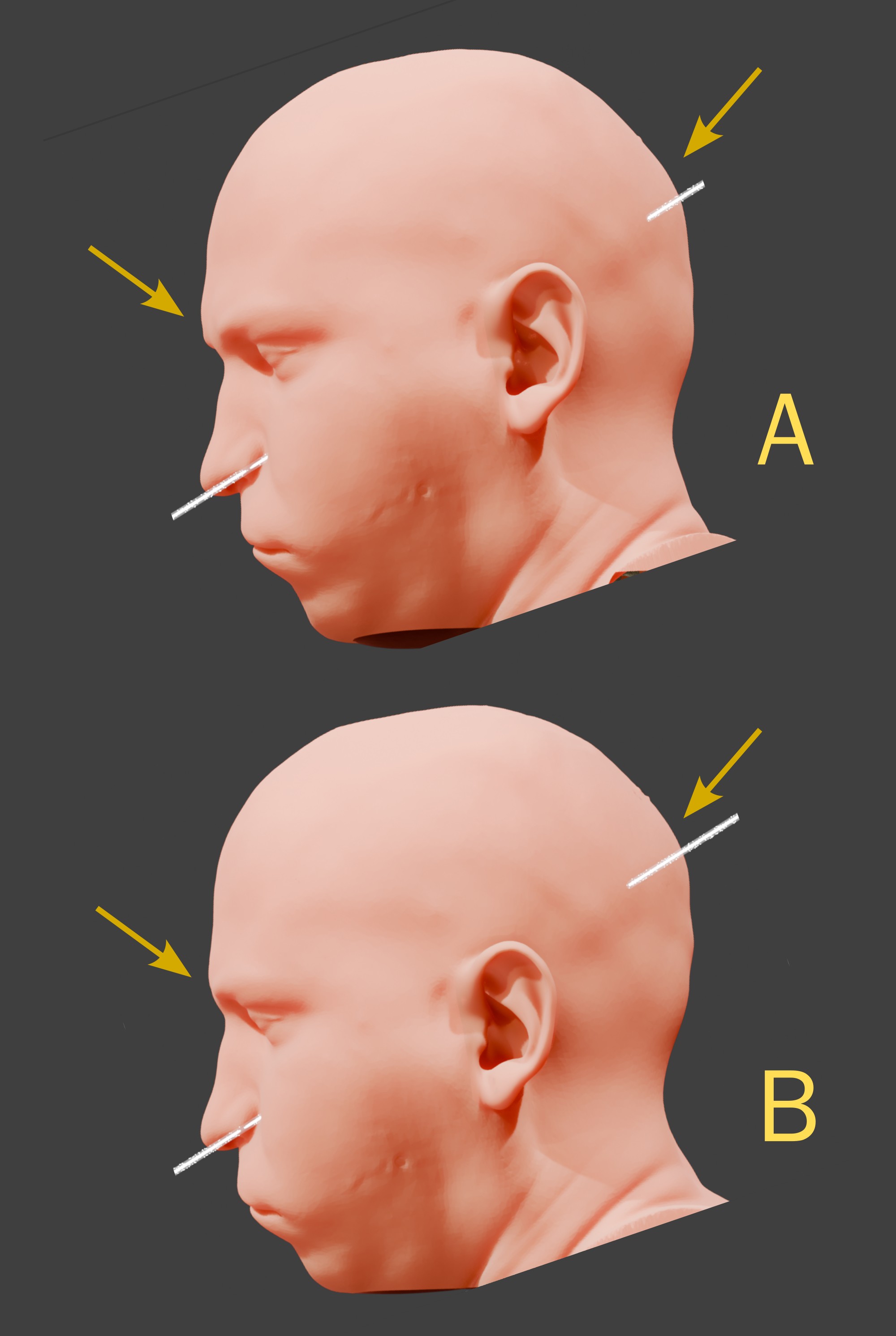
Fig. 3 A) Perspective of the official image, with a slightly angled profile view. B) True lateral view.
When observing the reconstructed head, with the proton beam following the injury trajectory, it can be verified that, from the perspective compatible with the image, i.e., the slightly angled profile (Fig. 3, A), the arrows indicate the eyebrow region and the beam entry point, located in the occipital region of the head. When compared to the true profile (Fig. 3, B), it is evident that the eyebrow is closer to the profile margin, while the beam entry point is further from the anterior limit.
For the reconstruction of the brain structure in voxel data [Moraes_et_al_2021_a], the tomography from [Edlow_et_al_2019_a] was used. For the reconstruction of the cerebral veins, an openly available examination for academic use was employed (https://data.kitware.com/#collection/591086ee8d777f16d01e0724/folder/58a372e38d777f0721a64dc6).
Results and Discussion
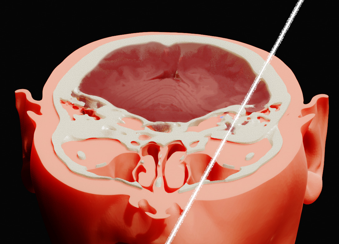
Fig. 4 Exact cut in the region traversed by the beam, according to the 3D reconstruction.
Upon performing the cut in the region traversed by the proton beam (Fig. 4), it is observed that the trajectory passed very close to the division between the occipital lobe and the temporal lobe, partially contradicting the most well-known image of the case, which indicates a clear entry in the occipital region, passing through the inferior temporal sulcus (https://img1.liveinternet.ru/images/attach/c/1//62/450/62450110_Trassa.jpg). The issue with the older image is that it suggests an entry almost at the center of the occipital region, which would be supported by the photograph. However, as demonstrated, the photograph was taken with a slight rotation, indicating that the entry point is potentially more displaced toward the left latero-lateral direction.
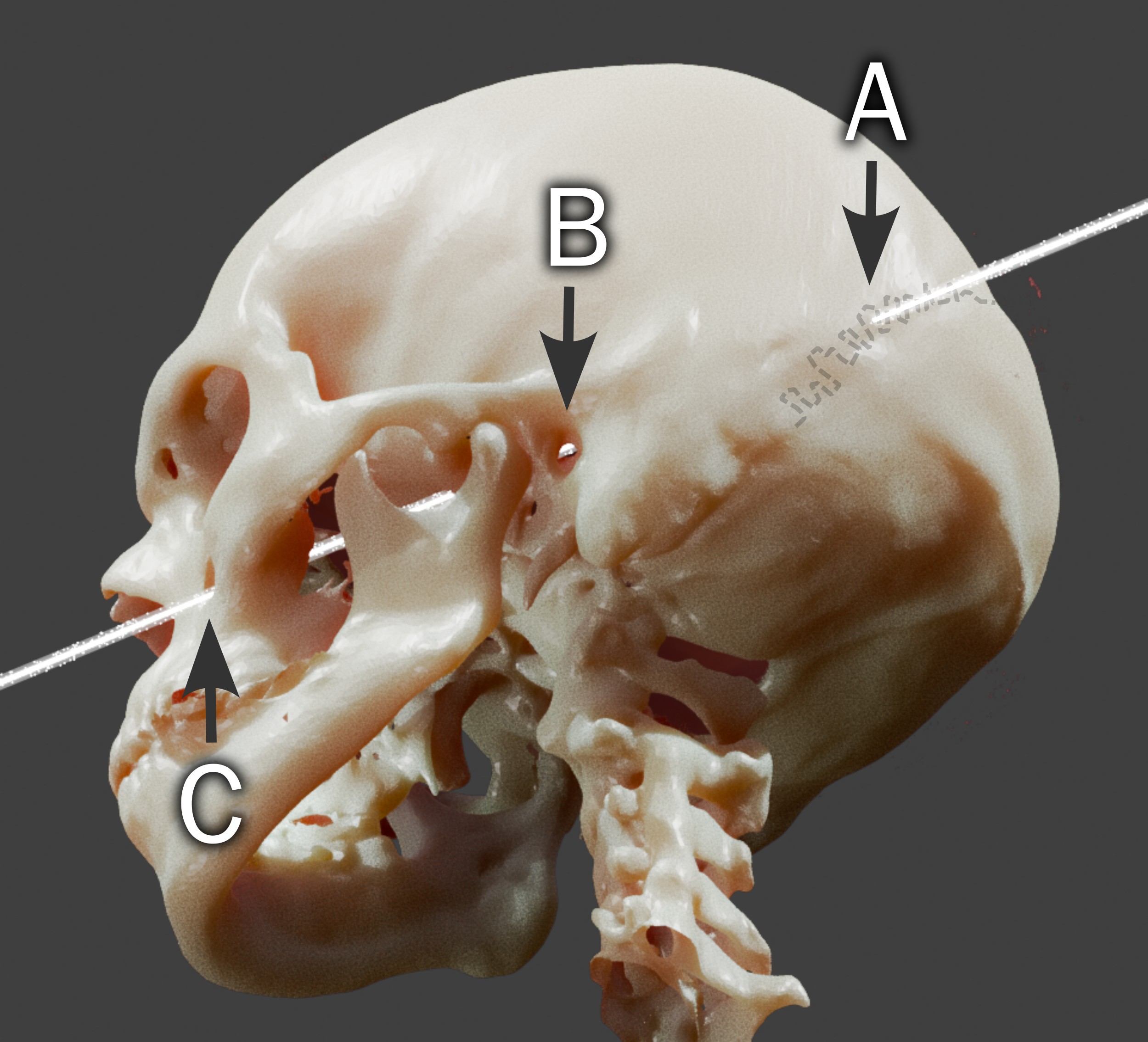
Fig. 5 Visualization of the skull and the beam’s path. A) Lambdoid suture. B) External acoustic meatus. C) Infraorbital foramen.
When observing the reconstructed skull laterally, three points are clearly distinguishable: the beam’s entry occurred in the transition region between the occipital bone and the left parietal bone, likely at the lambdoid suture (Fig. 5, A). The beam’s path can be identified from the left external acoustic meatus, in the epitympanic recess region (Fig. 5, B). The exit occurred at (or very close to) the left infraorbital foramen (Fig. 5, C).
An official medical description (according to a journalistic report) provides the following details:
“An intense high-energy proton beam, with transverse dimensions of 2 x 3 mm, followed the trajectory: occipital region of the head - mediobasal areas of the left temporal lobe - left temporal bone pyramid - bony labyrinth of the middle ear - tympanic cavity - mandibular fossa - tissues of the left nasal wing. The radiation dose at the entry was 200,000 roentgen, and at the exit, it was higher due to scattering in the material - 300,000 roentgen.” [Izvestia_1998_a]
It should be noted that, as this work is based on a lateral photograph, there is an inherent margin of error. However, the entry position is consistent with the description, as the beam technically strikes the occipital bone region, albeit at the boundary with the left parietal bone. The beam’s exit is also consistent with the description, but analyzing internal structures requires more specific observations.
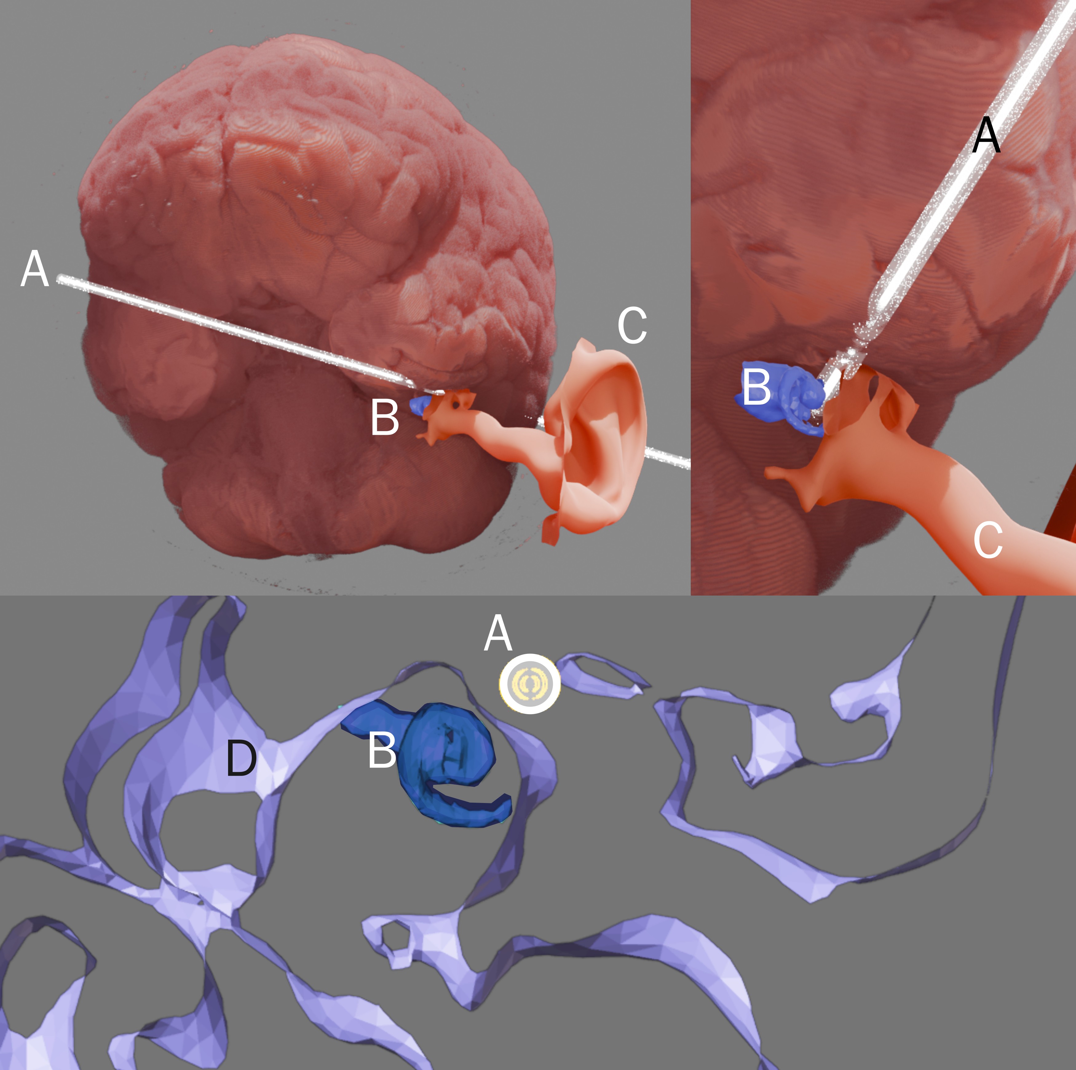
Fig. 6 Visualization of the proton beam in the inner ear region. A) Proton beam. B) Partial reconstruction of the bony labyrinth (limited by the resolution of the computed tomography). C) Surface mesh of soft tissue (internal acoustic meatus and ear). D) Surface mesh of bone tissue. The brain is the reddish-gray mass. The beam’s diameter is 3 mm.
The reconstructed proton beam trajectory (Fig. 6, A) passes very close to the bony labyrinth (Fig. 6, B), suggesting it may have struck the middle ear (Fig. 6, lower cut). Considering the margin of error, the trajectory is consistent with the medical description.
Regarding the cheek swelling, paresis, and hypoesthesia [Gessen_1997_a], a plausible explanation may lie in the potential injury to the infraorbital nerve. According to a study of 81 patients with this type of injury, 66.7% presented isolated hypoesthesia, with the cheek being the most affected area, at 42.8% [Devoti_et_al_2021_a]. In a case report involving infraorbital nerve injury, the patient reported numbness and pain in the cheek, nasal wing, and upper lip; post-treatment, the patient exhibited incomplete left infraorbital neuropathy and left facial neuropathy, consistent with the case under analysis [Lee_et_al_2020_a].
However, the trauma to the temporal bone, which caused hearing loss in Bugorski, may also have contributed to facial nerve paralysis. Delayed medical attention may have exacerbated the partial motor impairment, as patients treated promptly have a higher likelihood of recovery [Kurihara_et_al_2020_a].
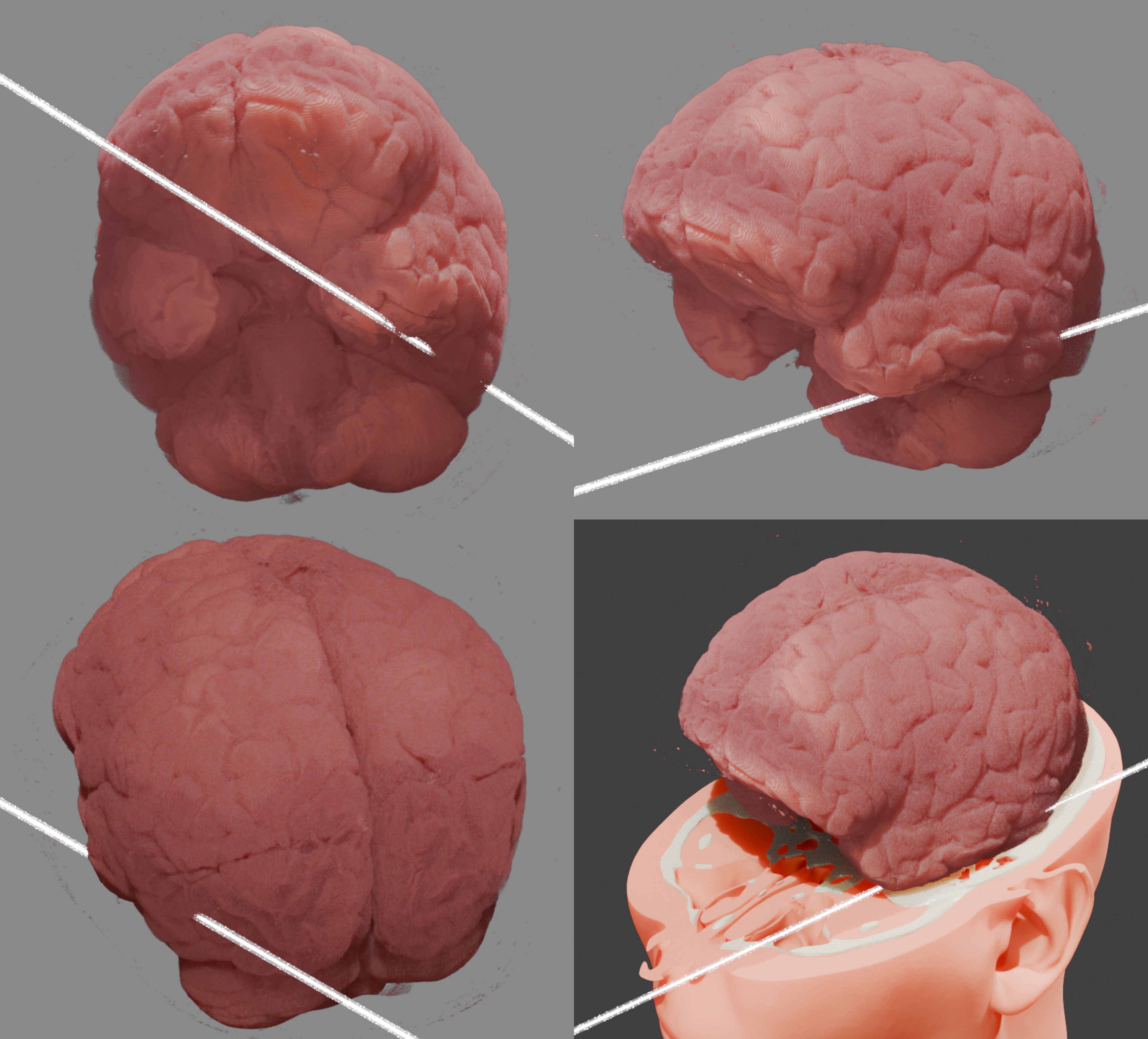
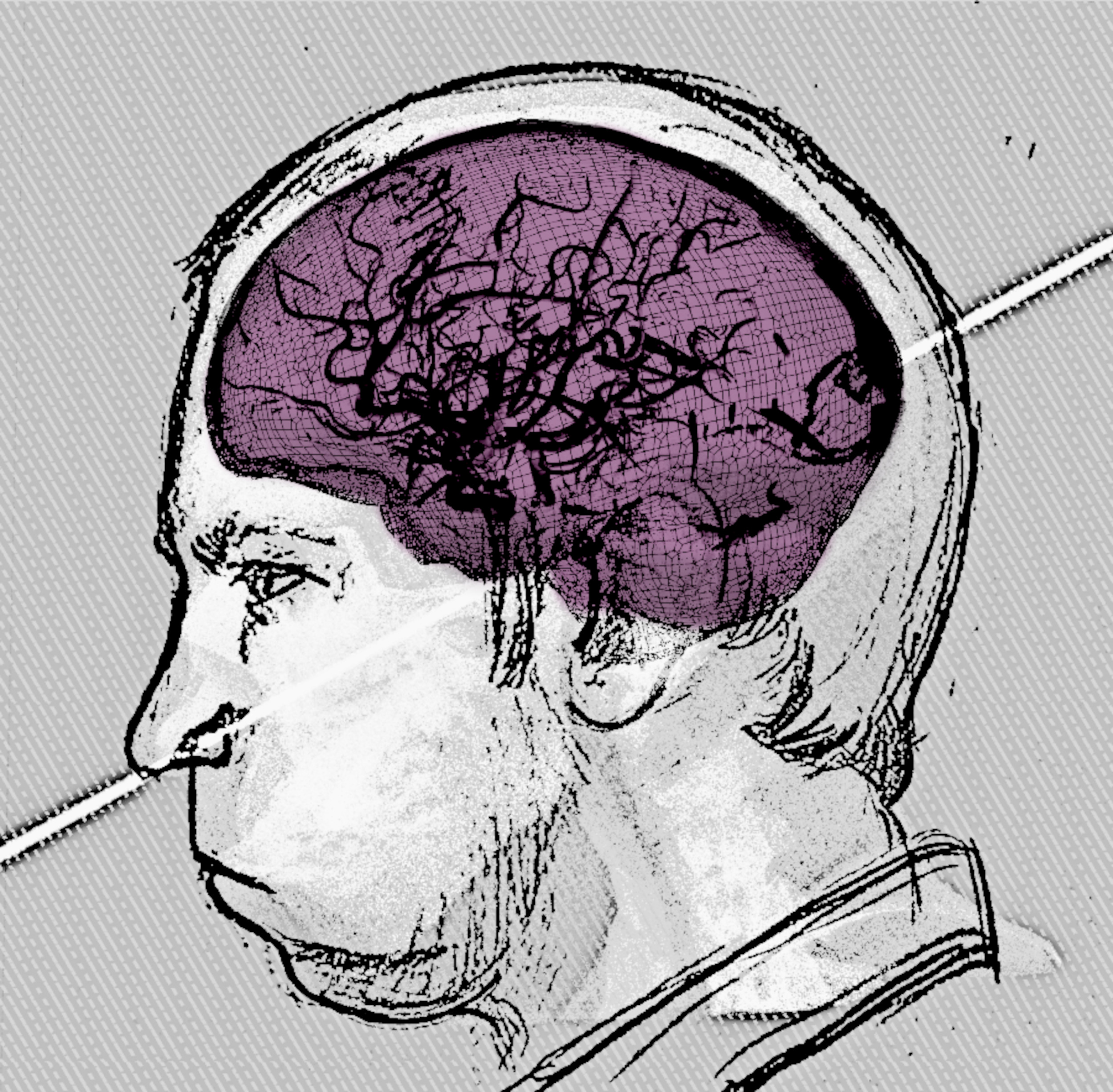
Fig. 7 Proton beam trajectory in relation to the brain.
Regarding the temporal region, specifically the temporal lobe, which is commonly associated with the development of epileptogenicity [Nayak_and_Bandyopadhyay_2023_a], it is observed that the majority of the proton beam struck this region, entering near the boundary with the occipital lobe, passing through the left temporal lobe, and exiting at or near the left inferior temporal sulcus (Fig. 7). According to the medical history, Bugorski experienced epilepsy and frequent seizures following the accident [Gessen_1997_a] [Izvestia_1998_a], which is consistent with the literature.
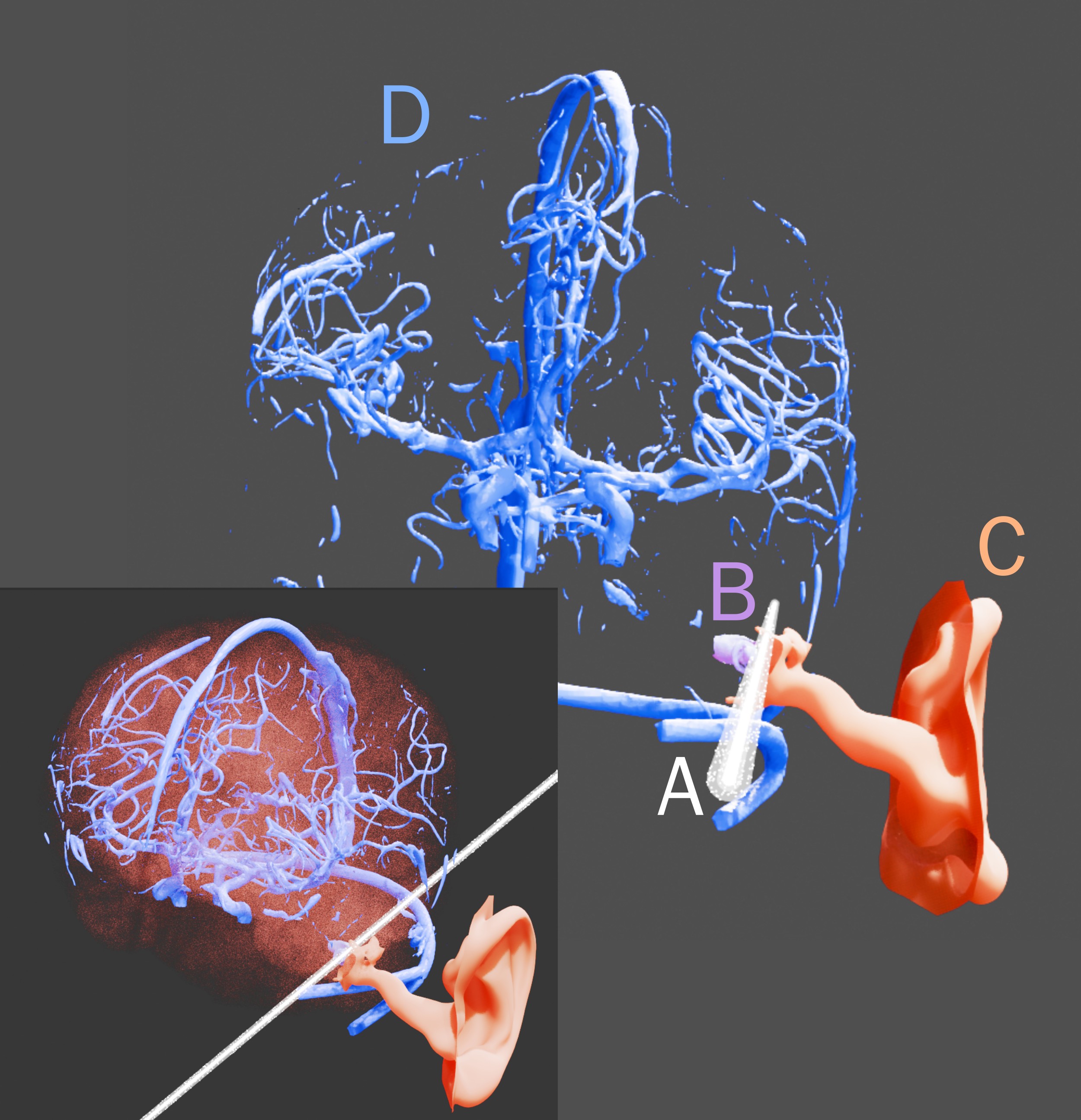
Fig. 8 Visualization of the proton beam in the region of superficial and deep cerebral veins. A) Proton beam. B) Partial reconstruction of the bony labyrinth. C) Surface mesh of soft tissue (internal acoustic meatus and ear). D) Superficial and deep cerebral veins (reconstruction limited by the resolution of the computed tomography). The brain is the transparent mass.
A reconstruction of the cerebral veins was performed to assess potential impact on significant circulatory structures. However, from the frontal observation aligned with the proton beam trajectory, there is no evidence of such an impact (Fig. 8, A and D).
Although the beam’s trajectory did not cause significant damage to the veins, it is documented that Bugorski experienced fatigue. Despite completing his kandidat nauk dissertation, he did not pursue a doktor nauk due to this condition, although he continued working at his post [Gessen_1997_a] [Arguments_and_Facts_2020_a]. This scenario aligns with the condition of fatigue and cognitive impairment following traumatic brain injury [Wright_et_al_2023_a].
Furthermore, it is noteworthy that, despite the injury occurring in a region associated with language (heard, spoken, and read) in the temporal lobe [Price_20212_a], and at the boundary with the occipital lobe, related to vision, the accident, apart from the expected fatigue, appears not to have significantly impaired Bugorski’s perception or intellectual faculties, as he continued working until at least 2020, at the age of 77. Despite the severity of the accident, factors such as his relatively young age (36 years), the small and well-defined injury area, and brain plasticity likely contributed to his recovery [Nieberlein_et_al_2023_a].
Study Limitations
Although all care was taken in the structural reconstruction process, this study is an approximation, and a margin of error must be taken into account, even with results consistent with documented medical data.
There is no pretension to establish a diagnosis, only to observe the academic literature regarding the injuries suffered by Bugorski in light of new studies, but given the anatomical complexity and variability of reactions from individual to individual, the findings present only some of a myriad of possibilities.
Having explained the situation, this study does not replace the original data, as well as the material and physical data of the event; however, as there is no documentation available, it represents a technical effort to elucidate the case, albeit subject to inaccuracies.
Conclusion
This study demonstrated the feasibility of a three-dimensional reconstruction of Anatoli Bugorski’s accident using tools such as OrtogOnBlender XP and virtual donor tomographies, providing a detailed analysis of the proton beam’s trajectory and its anatomical impacts. Despite limitations due to the scarcity of original documentation and reliance on low-resolution photographs, the results corroborate historical medical descriptions, elucidating injuries that explain symptoms such as hearing loss, epilepsy, and hypoesthesia. Brain plasticity and Bugorski’s relatively young age are factors that likely contributed to his partial recovery, allowing him to continue working until age 77. Beyond contributing to the understanding of this rare case, the work served as educational material, testing 3D modeling techniques applied to radiobiology. Future research could benefit from primary data, if available, to reduce the margin of error and deepen the analysis of radiation effects.
Acknowledgements
The authors express gratitude to Dr. Richard Gravalos, who provided the tomography of the virtual donor, enabling the reconstruction of the head and the didactic approach of this work.
References
Arguments and Facts. (2020, 17 de maio). «Ярче тысячи солнц». Невероятная история выжившего в синхротроне физика. aif.ru. https://aif.ru/society/science/yarche_tysyachi_solnc_neveroyatnaya_istoriya_vyzhivshego_v_sinhrotrone_fizika
Nayak, C. S., & Bandyopadhyay, S. (2023). Mesial Temporal Lobe Epilepsy. In StatPearls. StatPearls Publishing.. https://www.ncbi.nlm.nih.gov/books/NBK554432/
Devoti, J.-F., Nicot, R., Roland-Billecart, T., Ferri, J., & Schlund, M. (2021). Characterization of Infraorbital Nerve Sequelae After Orbital Floor or Zygomaticomaxillary Complex Fractures. Journal of Craniofacial Surgery, 33(1), 52–56. https://doi.org/10.1097/scs.0000000000007881
Edlow, B. L., Mareyam, A., Horn, A., Polimeni, J. R., Witzel, T., Tisdall, M. D., Augustinack, J., Stockmann, J. P., Diamond, B. R., Stevens, A., Tirrell, L. S., Folkerth, R. D., Wald, L. L., Fischl, B., & van der Kouwe, A. (2019). 7 Tesla MRI of the ex vivo human brain at 100 micron resolution. Cold Spring Harbor Laboratory. https://doi.org/10.1101/649822
Gessen, M. (1997, 1 de dezembro). The future ruins of the nuclear age. Wired. https://www.wired.com/1997/12/science-2/
Izvestia (1998). ПЕРСОНАЛЬНЫЙ ЧЕРНОБЫЛЬ АНАТОЛИЯ БУГОРСКОГО. Izvestia. https://www.liveinternet.ru/users/rewiever/post132062905
Kurihara, Y. Y., Fujikawa, A., Tachizawa, N., Takaya, M., Ikeda, H., & Starkey, J. (2020). Temporal Bone Trauma: Typical CT and MRI Appearances and Important Points for Evaluation. RadioGraphics, 40(4), 1148–1162. https://doi.org/10.1148/rg.2020190023
Lee, S. Y., Kim, S. H., Hwang, J. H., & Kim, K. S. (2020). Sensory recovery after infraorbital nerve avulsion injury. Archives of craniofacial surgery, 21(4), 244–248. https://doi.org/10.7181/acfs.2020.00290
Moraes, C., Graf, M., Dornelles, R., & Rosa, E. D. (2021). Reconstrução de Voxel Data no OrtogOnBlender. figshare. https://doi.org/10.6084/M9.FIGSHARE.13670134
Moraes, C., Startek, B., Dakir, I., Schreiner, T., Dornelles, R., & Rosa, E. da. (2025). Sistema Unificado para a Criação de um Crânio Composto no OrtogOnBlender XP. figshare. https://doi.org/10.6084/M9.FIGSHARE.28135760
msk1.ru. (2024, 16 de março). Пришла на выборы подругой, а ушла невестой. Житель Подмосковья сделал предложение своей девушке на избирательном участке. Публикуем трогательное видео и рассказываем, как это было. https://msk1.ru/text/gorod/2024/03/16/73343696/
Nieberlein, L., Rampp, S., Gussew, A., Prell, J., & Hartwigsen, G. (2023). Reorganization and Plasticity of the Language Network in Patients with Cerebral Gliomas. NeuroImage: Clinical, 37, 103326. https://doi.org/10.1016/j.nicl.2023.103326
Price, C. J. (2012). A review and synthesis of the first 20years of PET and fMRI studies of heard speech, spoken language and reading. NeuroImage, 62(2), 816–847. https://doi.org/10.1016/j.neuroimage.2012.04.062
Tank, P. W., & Gest, T. R. (2009). Atlas de Anatomia Humana. Artmed.
Wright, T. J., Elliott, T. R., Randolph, K. M., Pyles, R. B., Masel, B. E., Urban, R. J., & Sheffield-Moore, M. (2024). Prevalence of fatigue and cognitive impairment after traumatic brain injury. PLOS ONE, 19(3), e0300910. https://doi.org/10.1371/journal.pone.0300910